Fundamentals of Ionizing Radiation
From the Radiation imaging and science curriculum
TL;DR
Ionizing radiation is energy powerful enough to remove electrons from atoms, creating ions that can damage biological tissue. It comes from natural and man-made sources, categorized as either particulate or electromagnetic. Understanding its types and properties is crucial for safe and effective use in imaging and other applications.
1. The Mental Model
Think of ionizing radiation as tiny, energetic bullets or waves that can "knock off" electrons from atoms. When an atom loses an electron, it becomes an ion, which is chemically unstable and can react in ways that damage living cells.
2. The Core Material
Ionizing radiation is any type of particle or electromagnetic wave that carries enough energy to ionize an atom or molecule, meaning it can completely remove an electron from its orbit. This process changes the atom's chemical properties and can lead to cellular damage.
There are two main categories of ionizing radiation:
Particulate Radiation

Photo by PURPLE24 on Pexels
This type consists of tiny, fast-moving particles that have mass and carry kinetic energy. They directly interact with atoms, causing ionization.
- Alpha particles (α): These are essentially helium nuclei (2 protons, 2 neutrons). They are relatively heavy and carry a positive charge (+2). Because of their size and charge, they interact strongly with matter and lose energy quickly, meaning they have very short ranges and are easily stopped (e.g., by a sheet of paper or the outer layer of skin). However, if ingested or inhaled, they can cause significant localized damage.
- Beta particles (β): These are high-energy electrons (β-) or positrons (β+). They are much lighter and smaller than alpha particles, carry a single charge (-1 for electrons, +1 for positrons), and interact less strongly with matter. They have longer ranges than alpha particles and can penetrate a few millimeters to centimeters of tissue. They are stopped by materials like plastic or aluminum.
- Neutrons (n): These are uncharged particles found in the nucleus. Because they have no charge, they don't interact electromagnetically with electrons directly. Instead, they interact primarily with atomic nuclei, causing other particles to be emitted or making the nucleus unstable. They are highly penetrating and require thick, hydrogen-rich materials like water or concrete for shielding.
Electromagnetic Radiation
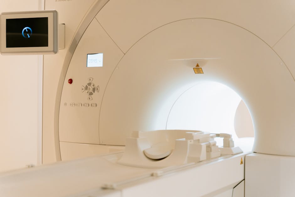
Photo by MART PRODUCTION on Pexels
This type consists of energy packets (photons) that have no mass and travel at the speed of light. They are part of the electromagnetic spectrum but possess enough energy to cause ionization.
- X-rays: Produced by accelerating electrons into a metal target. They are used extensively in medical imaging. Their energy levels vary, determining their penetrating power.
- Gamma rays (γ): Emitted from the nucleus of unstable (radioactive) atoms during radioactive decay. They are identical in nature to X-rays but typically have higher energies.
Key Interactions with Matter

Photo by Bastian Riccardi on Pexels
Understanding how different types of radiation interact with matter is fundamental to radiation protection and imaging.
graph TD
    A["Ionizing Radiation"] --> B{"Interacts with Matter"};
    B --> C["Particulate Radiation"];
    B --> D["Electromagnetic Radiation (Photons)"];

    C --> C1["Direct Ionization (Alpha, Beta)"];
    C --> C2["Nuclear Interactions (Neutrons)"];

    D --> D1["Photoelectric Effect"];
    D --> D2["Compton Scattering"];
    D --> D3["Pair Production"];
    D1 -- "Low Energy" --> E["Complete absorption, inner shell electron ejection"];
    D2 -- "Moderate Energy" --> F["Partial energy transfer, scattered photon, outer shell electron ejection"];
    D3 -- "High Energy (>1.02 MeV)" --> G["Photon converts to electron-positron pair"];
- Photoelectric Effect: A photon completely transfers its energy to an inner-shell electron, ejecting it from the atom. The photon ceases to exist. This is the primary interaction for X-rays in diagnostic imaging, especially in denser materials like bone, contributing to image contrast.
- Compton Scattering: A photon interacts with an outer-shell electron, imparting some of its energy to the electron and causing it to be ejected. The photon then scatters off at a different angle with reduced energy. This interaction is less desirable in imaging as it contributes to scatter radiation, which degrades image quality and increases patient/staff dose.
- Pair Production: A high-energy photon (energy > 1.02 MeV) interacts with the electric field of the nucleus, converting its entire energy into an electron-positron pair. This interaction is not relevant for diagnostic X-ray imaging but occurs with higher-energy gamma rays.
3. Worked Example
Let's compare the shielding requirements for different types of radiation.
Imagine you have three radioactive sources:
1. An alpha emitter
2. A beta emitter
3. A gamma emitter
You need to select appropriate materials to shield yourself from each.
- Alpha emitter: Since alpha particles have low penetration, a simple sheet of paper or even your skin's outer layer would effectively stop them. Your glove is sufficient.
- Beta emitter: Beta particles are more penetrating. You'd need something denser than paper, like a sheet of plastic (e.g., plexiglass, 1 cm thick) or a thin sheet of aluminum, to effectively attenuate them.
- Gamma emitter: Gamma rays are highly penetrating. To significantly reduce exposure, you'd need dense materials like lead or thick concrete. The thickness required depends on the energy of the gamma rays and the desired dose reduction. A few millimeters to several centimeters of lead might be necessary.
This example illustrates why different radiation types pose different safety challenges and require tailored protection strategies.
4. Key Takeaways
- Ionizing radiation has enough energy to remove electrons from atoms, creating ions.
- It's categorized into particulate radiation (alpha, beta, neutrons) and electromagnetic radiation (X-rays, gamma rays).
- Alpha particles are heavy, charged, and easily stopped but dangerous internally.
- Beta particles are lighter electrons/positrons with moderate penetration, stopped by plastic.
- Neutrons are uncharged and highly penetrating, requiring hydrogen-rich materials for shielding.
- X-rays and gamma rays are high-energy photons; they interact via the photoelectric effect, Compton scattering, and pair production.
- The photoelectric effect is key for imaging contrast; Compton scattering degrades image quality and increases dose.
- Different radiation types require different shielding materials and thicknesses.
Common Mistakes to Avoid:
- Assuming all radiation is equally dangerous or requires the same shielding.
- Confusing X-rays and gamma rays, as they originate differently but are electromagnetically identical.
- Underestimating the internal hazard of alpha emitters due to their low external penetrating power.
- Forgetting that Compton scattering contributes to both staff/patient dose and image degradation.
5. Now Try It
You're designing a radiation protection plan for a new imaging suite that uses X-rays. Think about the primary interactions of X-rays with the walls and staff, and briefly describe what materials you would recommend for the walls and why.
What to do:
1. Identify the main X-ray interaction(s) relevant to shielding the walls.
2. Suggest one or two common shielding materials.
3. Explain why those materials are effective based on your understanding of X-ray interactions.
What success looks like: You can clearly link the chosen shielding material to the specific X-ray interaction mechanisms, demonstrating an understanding of how they reduce radiation exposure.
Frequently asked about Fundamentals of Ionizing Radiation
Study this next
Get the full Radiation imaging and science curriculum
Clone the complete plan to your dashboard for unlimited AI-generated notes, practice quizzes, and a personalised revision schedule.
Save this course free