Overview of Eukaryotic Cell Structures
From the Biology curriculum
TL;DR
Eukaryotic cells are complex units of life, characterized by their true nucleus and membrane-bound organelles. These organelles perform specialized tasks, working together to maintain cell function and survival. Understanding their individual roles is key to grasping how a eukaryotic cell operates as a whole.
1. The Mental Model
Think of a eukaryotic cell as a tiny, bustling city. Each organelle is like a specific building or department with a unique job – power plant, factory, recycling center – all enclosed within a city wall (the cell membrane).
2. The Core Material
Eukaryotic cells are defined by their nucleus and other membrane-bound organelles. This internal compartmentalization allows for specialized functions to occur simultaneously and efficiently. Let's break down the main players:
The Nucleus: The Cell's Control Center
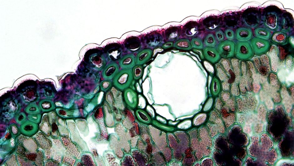
Photo by Fayette Reynolds M.S. on Pexels
The nucleus is usually the largest organelle and houses the cell's genetic material, DNA, organized into chromosomes. It's enclosed by a double membrane called the nuclear envelope, which has pores to regulate what enters and leaves. Inside, the nucleolus is where ribosomes are made. The nucleus controls cell growth, metabolism, and reproduction by directing protein synthesis.
Ribosomes: Protein Factories
Ribosomes are tiny organelles responsible for synthesizing proteins. They can be found free in the cytoplasm or attached to the endoplasmic reticulum. They read mRNA instructions to assemble amino acids into proteins.
Endoplasmic Reticulum (ER): Synthesis and Transport Highway

Photo by 隔壁光头老王 WangMing'Photo on Pexels
The endoplasmic reticulum (ER) is a vast network of interconnected membranes that extends throughout the cytoplasm. There are two types:
* Rough ER (RER): Studded with ribosomes, it's involved in synthesizing, folding, modifying, and transporting proteins destined for secretion or insertion into membranes.
* Smooth ER (SER): Lacks ribosomes and is involved in lipid synthesis, detoxification of drugs and poisons, and storage of calcium ions.
Golgi Apparatus: Packaging and Shipping Center

Photo by RDNE Stock project on Pexels
The Golgi apparatus (also called the Golgi complex or Golgi body) consists of flattened membrane-bound sacs called cisternae. It modifies, sorts, and packages proteins and lipids received from the ER into vesicles for secretion or delivery to other organelles.
Mitochondria: The Powerhouses
Mitochondria are often called the "powerhouses" of the cell because they are the primary sites of cellular respiration. This process converts glucose and oxygen into ATP, the cell's main energy currency. They have a double membrane, with the inner membrane folded into cristae to increase surface area for ATP production.
Lysosomes and Peroxisomes: Recycling and Detoxification
- Lysosomes: These spherical organelles contain powerful digestive enzymes. They break down waste materials, cellular debris, and foreign invaders (like bacteria), acting as the cell's recycling and waste disposal unit.
- Peroxisomes: Small, membrane-bound organelles that contain enzymes involved in metabolic reactions, particularly those that produce hydrogen peroxide (a toxic byproduct), which they then convert to water and oxygen. They're important for detoxifying harmful substances.
Vacuoles and Vesicles: Storage and Transport
- Vacuoles: Large, membrane-bound sacs primarily found in plant cells, where they store water, nutrients, and waste products, and help maintain turgor pressure. Animal cells can have small, temporary vacuoles.
- Vesicles: Smaller membrane-bound sacs used for transporting substances within the cell or for secreting them outside.
Cytoskeleton: Structure and Movement
The cytoskeleton is a network of protein filaments that provides structural support to the cell, helps maintain its shape, and plays a crucial role in cell movement, intracellular transport, and cell division. It's made of three main types of fibers: microtubules, microfilaments, and intermediate filaments.
Cell Membrane: The Border Patrol
The cell membrane (also called the plasma membrane) is the outer boundary of the animal cell, and it's just inside the cell wall in plant cells. It's a selective barrier made of a phospholipid bilayer with embedded proteins. It controls what enters and leaves the cell, mediates cell-to-cell communication, and maintains internal homeostasis.
Plant Cell Specifics: Cell Wall and Chloroplasts
Plant cells have a few extra structures:
* Cell Wall: A rigid outer layer made primarily of cellulose that provides structural support and protection to the cell.
* Chloroplasts: Sites of photosynthesis, where light energy is converted into chemical energy (glucose). They contain their own DNA and are typically green due to the presence of chlorophyll.
Here's how some key organelles work together for protein synthesis and transport:
graph TD
    A[Nucleus] --> B{Transcription};
    B --> C[mRNA];
    C --> D[Ribosome (on RER)];
    D --> E[Protein Synthesis];
    E --> F[Protein enters RER lumen];
    F --> G[Protein folding & modification];
    G --> H[Transport Vesicle];
    H --> I[Golgi Apparatus (cis face)];
    I --> J[Protein further modified & sorted];
    J --> K[Golgi Apparatus (trans face)];
    K --> L[Secretory Vesicle];
    L --> M[Cell Membrane];
    M --> N[Protein Secreted];
3. Worked Example
Imagine a pancreatic cell that needs to produce and secrete insulin, a protein hormone.
1. Nucleus: The gene for insulin is transcribed into mRNA.
2. Ribosomes (on RER): The mRNA travels to ribosomes attached to the Rough ER. Here, insulin protein is synthesized and directly enters the ER lumen.
3. Rough ER: Insulin folds into its correct 3D shape and might get some initial modifications.
4. Transport Vesicle: A small vesicle buds off from the ER, carrying the insulin.
5. Golgi Apparatus: The vesicle fuses with the cis face of the Golgi. Insulin then moves through the Golgi's cisternae, undergoing further modifications, sorting, and packaging.
6. Secretory Vesicle: At the trans face, insulin is packaged into a secretory vesicle.
7. Cell Membrane: This vesicle travels to the cell membrane, fuses with it, and releases the insulin outside the cell (exocytosis) into the bloodstream, where it can act on target cells.
4. Key Takeaways
- Eukaryotic cells are defined by a nucleus and membrane-bound organelles that specialize in different functions.
- The nucleus houses DNA and controls cellular activities.
- Mitochondria produce ATP for energy through cellular respiration.
- The ER and Golgi apparatus work together to synthesize, modify, and transport proteins and lipids.
- Lysosomes and peroxisomes are crucial for waste breakdown and detoxification.
- Plant cells have unique structures like a cell wall and chloroplasts for protection and photosynthesis.
- The cell membrane regulates passage of substances and facilitates cell communication.
Common mistakes to avoid:
- Confusing the functions of rough ER (protein synthesis/modification) and smooth ER (lipid synthesis/detoxification).
- Forgetting that ribosomes are responsible for protein synthesis, regardless of where they are located (free or on ER).
- Mixing up the roles of mitochondria (energy production) and chloroplasts (photosynthesis).
- Thinking that vacuoles are only in plant cells; animal cells can have small, temporary ones.
5. Now Try It
Take a blank piece of paper and sketch out a typical animal cell. Label at least 10 major organelles we discussed, and next to each label, write down one key function in your own words. Then, on the same paper, add the structures unique to a plant cell and their functions. You'll know you've got it when you can explain the core function of each labeled organelle without looking at your notes.
Frequently asked about Overview of Eukaryotic Cell Structures
More from Biology
Get the full Biology curriculum
Clone the complete plan to your dashboard for unlimited AI-generated notes, practice quizzes, and a personalised revision schedule.
Save this course free