Connective Tissue: Diversity and Support
From the Sports Med Unit Two curriculum
TL;DR
Connective tissues are crucial for supporting, connecting, and protecting your body, acting like the "glue" that holds everything together. They're incredibly diverse, ranging from loose tissues that cushion organs to dense tissues forming tendons and bones. Understanding their unique structures and functions helps you grasp how they contribute to overall body mechanics and injury resilience.
1. The Mental Model
Think of connective tissue as your body's internal scaffolding and packing material. It's everywhere, providing structure, connecting different parts, and filling spaces. It's not just one thing; instead, it's a family of tissues with different jobs, all built from similar basic components but arranged in unique ways to fit their specific roles.
2. The Core Material
Connective tissues (CT) are defined by three main components: specialized cells, protein fibers, and ground substance. It's the extracellular matrix (ECM) – the fibers and ground substance – that largely determines the tissue's properties. Unlike epithelial tissue, CT typically isn't directly exposed to the outside environment and usually has a good blood supply (except for cartilage).
Components of Connective Tissue

Photo by Leo Freire on Pexels
- Specialized Cells: These cells produce and maintain the ECM.
- Fibroblasts: The most common CT cells, they produce fibers and ground substance.
- Adipocytes: Fat cells that store energy.
- Chondrocytes: Cartilage cells.
- Osteocytes: Bone cells.
- Immune cells: Macrophages, mast cells, plasma cells that defend the body.
- Protein Fibers: Provide strength and elasticity.
- Collagen fibers: Strongest and most abundant, providing tensile strength (resistance to stretching). Think of them like steel cables.
- Elastic fibers: Stretchy and resilient, allowing tissues to return to their original shape after distortion. Think of them like rubber bands.
- Reticular fibers: Thin, branching fibers that form delicate networks, providing structural support to soft organs like the spleen.
- Ground Substance: The clear, viscous (or sometimes solid) material that fills the space between cells and fibers. It consists of water, proteoglycans, and glycoproteins, acting as a lubricant and barrier.
Types of Connective Tissue
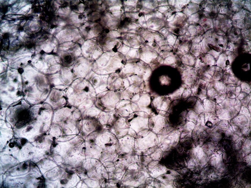
Photo by turek on Pexels
Connective tissues are broadly categorized into connective tissue proper and specialized connective tissue.
graph TD
    A["Connective Tissue"] --> B["Connective Tissue Proper"]
    A --> C["Specialized Connective Tissue"]

    B --> D["Loose CT"]
    B --> E["Dense CT"]

    D --> F["Areolar (Loose)"]
    D --> G["Adipose (Fat)"]
    D --> H["Reticular"]

    E --> I["Dense Regular"]
    E --> J["Dense Irregular"]
    E --> K["Elastic"]

    C --> L["Cartilage"]
    C --> M["Bone"]
    C --> N["Blood"]
    C --> O["Lymph"]

    L --> P["Hyaline"]
    L --> Q["Elastic"]
    L --> R["Fibrocartilage"]
- Connective Tissue Proper:
- Loose Connective Tissue: Has fewer fibers and more ground substance. It's flexible and acts as a packing material.
- Areolar: Most common; cushions organs, holds skin to underlying tissues. Contains all fiber types.
- Adipose: Fat storage, insulation, shock absorption. Mostly adipocytes.
- Reticular: Forms soft internal skeleton for organs like the spleen, lymph nodes, and bone marrow.
- Dense Connective Tissue: Has many fibers and less ground substance, providing strength.
- Dense Regular: Collagen fibers are arranged parallel, providing high tensile strength in one direction. Found in tendons (muscle to bone) and ligaments (bone to bone).
- Dense Irregular: Collagen fibers are irregularly arranged, providing strength in multiple directions. Found in the dermis of the skin, organ capsules.
- Elastic: Dominated by elastic fibers, allowing stretch and recoil. Found in large artery walls, vocal cords.
- Loose Connective Tissue: Has fewer fibers and more ground substance. It's flexible and acts as a packing material.
- Specialized Connective Tissue:
- Cartilage: Firm but flexible, avascular (no blood supply), heals slowly.
- Hyaline: Most common; reduces friction in joints (articular cartilage), forms ribs, nose, trachea.
- Elastic: Flexible support; ear, epiglottis.
- Fibrocartilage: Strongest, resists compression; intervertebral discs, menisci of knee.
- Bone: Hard, rigid tissue for support, protection, and mineral storage.
- Blood & Lymph: Fluid connective tissues for transport.
- Cartilage: Firm but flexible, avascular (no blood supply), heals slowly.
3. Worked Example
Imagine you're examining a cross-section of a tendon. What would you expect to see?
A tendon's job is to connect muscle to bone and transmit force, meaning it needs to be incredibly strong and resist stretching along its length. Therefore, you'd expect to see:
- Cells: Primarily fibroblasts (or fibrocytes, their mature form) which are long, thin cells scattered between the fibers. Their job is to produce and maintain the matrix.
- Fibers: An abundance of densely packed, parallel collagen fibers. These fibers are arranged in this highly organized way to provide maximum tensile strength in the direction of muscle pull. You wouldn't see many elastic or reticular fibers, as elasticity isn't its primary function, and delicate networks aren't needed.
- Ground Substance: Very little ground substance. The space is mostly filled with the tightly packed collagen fibers.
This specific arrangement of collagen fibers (dense, parallel) and sparse cells with minimal ground substance is characteristic of dense regular connective tissue, perfectly suited for the tendon's function.
4. Key Takeaways
- Connective tissues are broadly classified based on their cell types, fiber composition, and ground substance.
- The extracellular matrix (fibers + ground substance) largely dictates the specific properties and functions of each connective tissue type.
- Collagen fibers provide strength and resistance to pulling forces, while elastic fibers allow for stretch and recoil.
- Tendons and ligaments are examples of dense regular connective tissue, designed for high tensile strength in one direction.
-
Cartilage is a unique connective tissue because it's avascular, relying on diffusion for nutrients, which affects its repair capabilities.
-
Common Mistakes:
- Confusing tendons (muscle to bone) with ligaments (bone to bone); both are dense regular CT but have different attachments.
- Thinking all connective tissue is dense and strong; much of it is loose and acts as filler.
- Forgetting that blood is also a connective tissue, even though it's fluid.
- Assuming all connective tissues have a rich blood supply; cartilage is a key exception.
5. Now Try It
Choose three different locations in the body (e.g., knee joint, outer ear, underneath your skin) and identify the primary type(s) of connective tissue you'd find there. For each location, briefly explain why that specific connective tissue is well-suited for its function in that area, referencing its cell types, fiber arrangement, and ground substance characteristics. Success looks like you accurately identifying the CT types and clearly explaining the functional advantage of their specific composition for each location.
Frequently asked about Connective Tissue: Diversity and Support
More from Sports Med Unit Two
Get the full Sports Med Unit Two curriculum
Clone the complete plan to your dashboard for unlimited AI-generated notes, practice quizzes, and a personalised revision schedule.
Save this course free