Muscle Tissue: Movement and Contraction
From the Sports Med Unit Two curriculum
TL;DR
Muscle tissue is specialized for contraction, allowing for movement, maintaining posture, and producing heat. This contraction happens through the sliding filament model, where actin and myosin proteins interact. The nervous system controls muscle activity, determining when and how strongly muscles contract.
1. The Mental Model
Think of your muscles like tiny, super-efficient ropes made of millions of even tinier threads. When your brain sends a signal, these threads pull on each other, making the rope shorter and thicker, which moves your bones.
2. The Core Material
Muscle tissue is one of the four main tissue types in your body, and its primary job is to generate force and movement. There are three main types of muscle tissue, each with distinct roles:
Skeletal Muscle
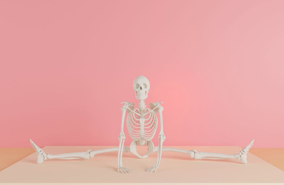
Photo by dada _design on Pexels
Skeletal muscle is what you typically think of when you hear "muscle." It's attached to bones and is responsible for voluntary movements like walking, lifting, and running. These muscles are striated, meaning they have a striped appearance under a microscope due to the organized arrangement of contractile proteins.
Cardiac Muscle

Photo by Mohammad Ubaid on Pexels
Cardiac muscle is found only in the walls of your heart. It's also striated but is involuntary, meaning you don't consciously control its contractions. Its continuous, rhythmic pumping action is essential for circulating blood throughout your body.
Smooth Muscle
Photo by Mohammad Ubaid on Pexels
Smooth muscle is found in the walls of internal organs like your stomach, intestines, and blood vessels. It's non-striated and involuntary, responsible for slow, sustained contractions, such as moving food through your digestive tract or regulating blood pressure.
The Sliding Filament Model of Contraction
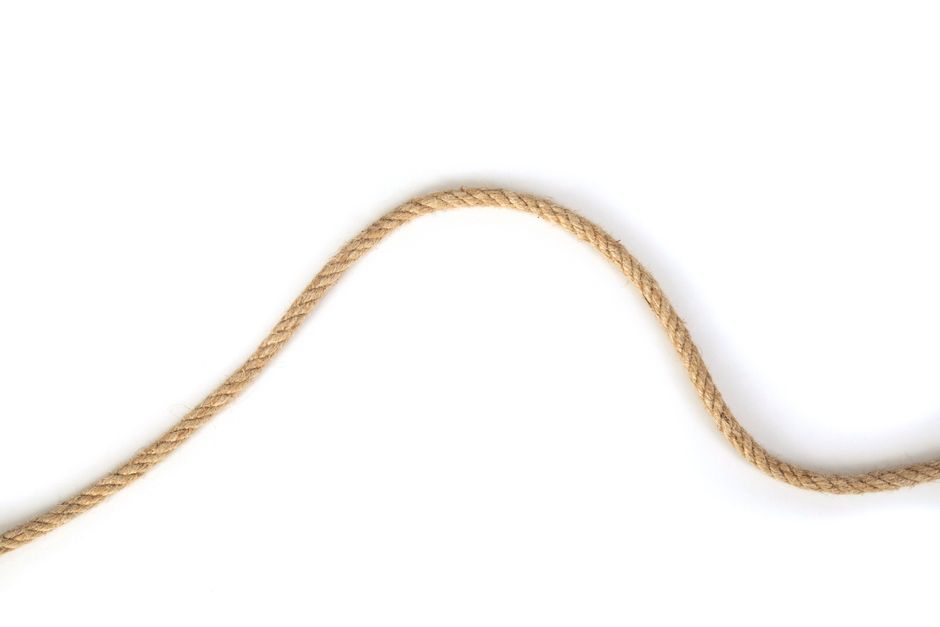
Photo by Engin Akyurt on Pexels
Muscle contraction, especially in skeletal and cardiac muscle, is explained by the sliding filament model. This model describes how thin filaments (made primarily of the protein actin) slide past thick filaments (made primarily of the protein myosin), causing the muscle fiber to shorten.
Here's the simplified process:
1. Nerve Impulse: A signal from your brain (or spinal cord) reaches the muscle fiber.
2. Calcium Release: This signal triggers the release of calcium ions within the muscle cell.
3. Myosin Binding: Calcium allows myosin heads to bind to actin, forming cross-bridges.
4. Power Stroke: The myosin heads pivot, pulling the actin filaments towards the center of the sarcomere (the basic contractile unit of a muscle fiber). This is like a tiny rowing motion.
5. ATP Detachment: A molecule of ATP (adenosine triphosphate, your body's energy currency) binds to the myosin head, causing it to detach from actin.
6. ATP Hydrolysis & Re-cocking: ATP is broken down into ADP and phosphate, providing energy for the myosin head to "re-cock" or return to its original position, ready to bind again.
7. Repetition: This cycle of binding, pulling, detaching, and re-cocking repeats as long as calcium and ATP are available, causing continuous shortening of the muscle.
graph TD
    A["Nerve Impulse Reaches Muscle"] --> B["Acetylcholine Release (Neurotransmitter)"];
    B --> C["Muscle Cell Membrane Depolarizes"];
    C --> D["Calcium Ions Released from Sarcoplasmic Reticulum"];
    D --> E["Calcium Binds to Troponin"];
    E --> F["Tropomyosin Shifts, Exposing Actin Binding Sites"];
    F --> G["Myosin Heads Bind to Actin (Cross-Bridge Formation)"];
    G --> H["Power Stroke (Myosin Pulls Actin)"];
    H --> I["ATP Binds to Myosin Head"];
    I --> J["Myosin Detaches from Actin"];
    J --> K["ATP Hydrolyzed, Myosin Head 'Re-cocks'"];
    K --> L{"Is Calcium Still Present?"};
    L -- Yes --> G;
    L -- No --> M["Calcium Pumped Back into Sarcoplasmic Reticulum"];
    M --> N["Tropomyosin Re-covers Actin Binding Sites"];
    N --> O["Muscle Relaxes"];
3. Worked Example
Imagine you're lifting a dumbbell. Your brain sends a signal down your spinal cord to the motor neurons connected to your biceps muscle. At the neuromuscular junction, the neurotransmitter acetylcholine is released, sparking an electrical impulse in your bicep muscle fibers. This impulse travels deep into the muscle, causing calcium to flood out from storage areas. These calcium ions then bind to proteins on the actin filaments, moving another protein (tropomyosin) out of the way. Now, the myosin heads on the thick filaments can grab onto the exposed binding sites on the actin. With energy from ATP, these myosin heads pivot, pulling the actin filaments inwards, shortening the muscle. This shortening generates the force needed to lift the dumbbell. When you put the dumbbell down, the nerve signal stops, calcium is pumped away, and the muscle relaxes.
4. Key Takeaways
- Muscle tissue allows movement, posture maintenance, and heat generation.
- There are three types of muscle: skeletal (voluntary, striated), cardiac (involuntary, striated), and smooth (involuntary, non-striated).
- Muscle contraction fundamentally relies on the sliding filament model, where actin and myosin proteins interact.
- Calcium ions play a critical role by unblocking binding sites for myosin on actin.
- ATP provides the energy for myosin heads to detach, re-cock, and perform the power stroke.
- Nerve impulses initiate the entire contraction process by triggering calcium release.
Common mistakes to avoid:
- Confusing the roles of actin and myosin; myosin is the "puller," actin is the "pulled."
- Forgetting that ATP is essential not just for the power stroke but also for detaching myosin from actin.
- Thinking all muscle contractions are voluntary; cardiac and smooth muscles are involuntary.
- Assuming calcium is only important for bone health; it's crucial for muscle contraction too.
5. Now Try It
Spend 15 minutes drawing a simple diagram of a sarcomere (the basic contractile unit), showing the thick (myosin) and thin (actin) filaments. Then, mentally trace the path of a nerve signal causing contraction, labeling where calcium and ATP would be involved in your diagram. When you're done, check if your drawing clearly shows how the filaments would slide past each other during contraction.
Frequently asked about Muscle Tissue: Movement and Contraction
More from Sports Med Unit Two
Get the full Sports Med Unit Two curriculum
Clone the complete plan to your dashboard for unlimited AI-generated notes, practice quizzes, and a personalised revision schedule.
Save this course free