Nervous Tissue: Communication and Control
From the Sports Med Unit Two curriculum
TL;DR
Your nervous system uses specialized cells called neurons to send electrical and chemical signals throughout your body, allowing for rapid communication and control. These signals travel along axons, jump across synapses to other neurons or target cells, and enable everything from movement to thought. Understanding this intricate network is key to comprehending how your body responds to stimuli and performs actions.
1. The Mental Model
Think of your nervous system as your body's internet: a vast network of wires (nerves) and processing centers (brain, spinal cord) that instantly sends messages. These messages are like tiny electrical pulses and chemical signals, allowing different parts of your body to talk to each other and coordinate actions.
2. The Core Material
Your nervous system is made of nervous tissue, which contains two main types of cells: neurons and neuroglia (or glial cells). Neurons are the communicators, sending signals, while neuroglia support and protect them.
Neuron Structure and Function
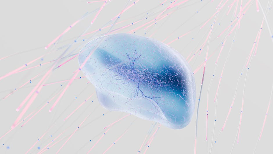
Photo by Google DeepMind on Pexels
A typical neuron has three main parts:
- Dendrites: These are tree-like branches that receive signals from other neurons. Think of them as the antenna.
- Cell Body (Soma): This is the neuron's "control center," containing the nucleus and other organelles. It processes incoming signals.
- Axon: A long, slender extension that transmits electrical signals (action potentials) away from the cell body to other neurons or target cells (like muscles or glands). Imagine this as the cable.
Many axons are covered in a myelin sheath, a fatty layer that insulates the axon and speeds up signal transmission. Gaps in the myelin sheath are called Nodes of Ranvier, where the signal "jumps" from one node to the next.
How Neurons Communicate: Action Potentials and Synapses
Photo by Google DeepMind on Pexels
Neurons communicate using electrical signals (action potentials) and chemical signals (neurotransmitters).
-
Action Potential (Electrical Signal): This is a brief, rapid change in the electrical potential across the neuron's membrane. When a neuron receives enough stimulation, it reaches a threshold, triggering an action potential. This signal travels down the axon.
- Resting Membrane Potential: A neuron at rest has a slightly negative charge inside compared to the outside, maintained by the sodium-potassium pump.
- Depolarization: When a stimulus arrives, sodium (Na+) channels open, and Na+ rushes into the cell, making the inside positive.
- Repolarization: Potassium (K+) channels open, and K+ rushes out, making the inside negative again.
- Refractory Period: A brief time when the neuron can't fire another action potential, ensuring one-way signal flow.
-
Synaptic Transmission (Chemical Signal): When the action potential reaches the end of the axon (the axon terminal), it can't directly jump to the next neuron. Instead, it triggers the release of neurotransmitters into the synaptic cleft (the tiny gap between neurons).
- Neurotransmitters bind to receptors on the postsynaptic neuron's dendrite or cell body, opening ion channels and either exciting or inhibiting the next neuron.
- This chemical signal is quickly cleared from the synapse (e.g., reabsorbed or broken down) to allow for new signals.
Here's a simplified flow of neural communication:
graph TD
    A["Stimulus Received by Dendrite"] --> B["Cell Body Integrates Signal"]
    B --> C{Reach Threshold?}
    C -- No --> A
    C -- Yes --> D["Action Potential Generated"]
    D --> E["Action Potential Travels down Axon"]
    E --> F["Axon Terminal Reached"]
    F --> G["Neurotransmitter Release into Synaptic Cleft"]
    G --> H["Neurotransmitter Binds to Postsynaptic Receptors"]
    H --> I{"Postsynaptic Cell Responds (Excitation/Inhibition)"}
    I -- Excitation --> D_Next["Generate Action Potential (Next Neuron)"]
    I -- Inhibition --> A_Next["Prevent Action Potential (Next Neuron)"]
Neuroglia (Glial Cells)
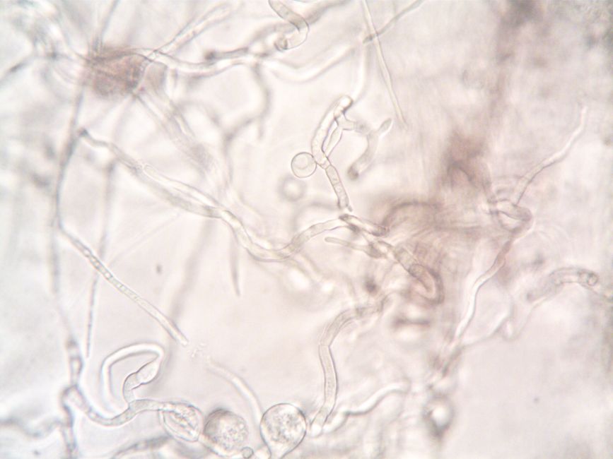
Photo by turek on Pexels
These support cells are crucial for neuron function. Key types include:
- Astrocytes: Provide nutrients, maintain ion balance, and form the blood-brain barrier.
- Oligodendrocytes (CNS) & Schwann Cells (PNS): Produce myelin sheaths around axons, speeding up signal conduction.
- Microglia: Immune cells of the nervous system, clearing debris and pathogens.
3. Worked Example
Imagine you accidentally touch a hot stove.
- Stimulus: Heat receptors in your fingertips detect the high temperature.
- Sensory Neuron Activation: These receptors generate an electrical signal (action potential) in a sensory neuron.
- Signal Transmission: The action potential travels rapidly along the sensory neuron's axon, up your arm, and into your spinal cord.
- Synaptic Transmission (Spinal Cord): At a synapse in the spinal cord, the sensory neuron releases neurotransmitters. These bind to receptors on an interneuron.
- Interneuron Processing: The interneuron quickly relays the signal to a motor neuron.
- Synaptic Transmission (Motor Neuron): The motor neuron generates its own action potential, which travels down its axon out of the spinal cord and to the muscles in your arm.
- Neuromuscular Junction: At the end of the motor neuron's axon (at the neuromuscular junction), it releases a specific neurotransmitter (acetylcholine).
- Muscle Contraction: Acetylcholine binds to receptors on your arm muscles, causing them to contract, and you rapidly pull your hand away from the stove. All of this happens in a fraction of a second!
4. Key Takeaways
- Neurons are specialized cells for transmitting electrical and chemical signals throughout your body.
- Dendrites receive signals, the cell body processes them, and the axon transmits action potentials.
- The myelin sheath insulates axons and dramatically increases the speed of signal conduction.
- Action potentials are electrical signals caused by rapid changes in ion flow across the neuron's membrane.
- Synapses are the junctions where neurons communicate chemically using neurotransmitters.
- Neuroglia are crucial support cells that protect neurons, provide nutrients, and form myelin.
- The rapid, coordinated communication via nervous tissue allows for swift responses to stimuli.
Common Mistakes to Avoid:
- Thinking signals jump directly from neuron to neuron without a synapse.
- Confusing the role of dendrites (receive) with axons (transmit).
- Underestimating the importance of neuroglia; they're not just passive support.
- Forgetting that an action potential is an electrical event, while synaptic transmission is chemical.
5. Now Try It
For 15 minutes, draw and label a detailed diagram of a neuron. Include the dendrites, cell body, nucleus, axon, myelin sheath, Nodes of Ranvier, and axon terminals. Then, using arrows, illustrate the general direction of signal flow through the neuron. Below your diagram, write a short paragraph explaining how myelin contributes to the speed of signal transmission.
Success looks like a clearly labeled diagram with correct signal flow, and an explanation that accurately describes how myelin improves conduction velocity by allowing the action potential to jump between Nodes of Ranvier.
Frequently asked about Nervous Tissue: Communication and Control
More from Sports Med Unit Two
Get the full Sports Med Unit Two curriculum
Clone the complete plan to your dashboard for unlimited AI-generated notes, practice quizzes, and a personalised revision schedule.
Save this course free