Organs: Coordinated Tissue Function
From the biolgyu curriculum
TL;DR
Organs are biological structures made of multiple types of tissues working together to perform specific functions. Different tissues within an organ cooperate through specialized cells and intercellular communication. This coordinated effort allows organs to carry out complex tasks essential for an organism's survival.
1. The Mental Model
Think of an organ like a specialized team, where each team member (tissue type) has a unique role, but they all work together toward a common goal. No single tissue can do the organ's job alone; they all depend on each other.
2. The Core Material
You know that cells are the basic units of life, and similar cells often group to form tissues (like muscle tissue or nervous tissue). When different types of tissues come together to perform a specific, larger job, they form an organ.
For an organ to function, these different tissues don't just sit next to each other; they actively coordinate their activities. This coordination happens through several mechanisms:
a. Structural Arrangement

Photo by Talha Kuğu on Pexels
Tissues are physically arranged in specific ways within an organ. For example, in the stomach, epithelial tissue lines the interior, smooth muscle tissue provides contractions, connective tissue holds it all together, and nervous tissue controls these actions. Their positions are crucial for their functions.
b. Intercellular Communication
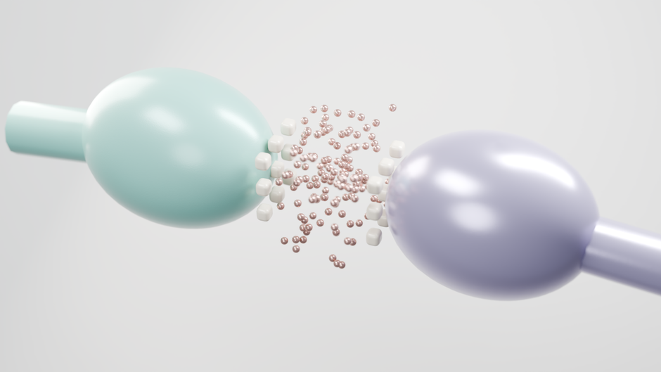
Photo by Santhosh Kanthala on Pexels
Cells within and between different tissues communicate constantly. This can happen via:
* Chemical signals: Hormones or neurotransmitters released by one cell affecting another.
* Direct contact: Cells touching each other, sharing information through specialized junctions.
* Electrical signals: Rapid communication, especially important in nervous and muscle tissues.
c. Functional Specialization
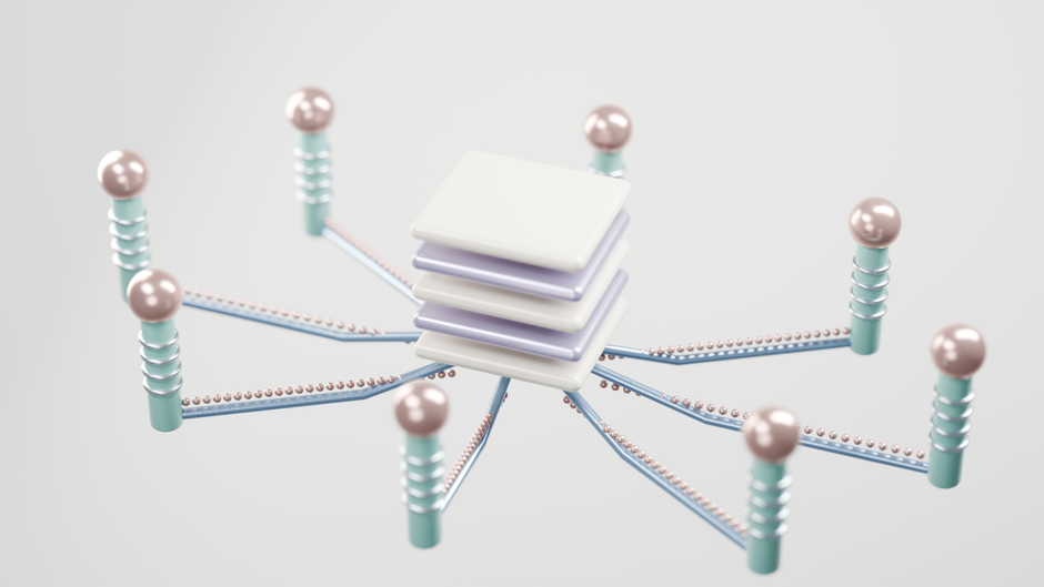
Photo by Santhosh Kanthala on Pexels
Each tissue type has specialized cells designed for particular tasks.
* Epithelial tissue: Protection, secretion, absorption.
* Connective tissue: Support, binding, transport.
* Muscle tissue: Movement.
* Nervous tissue: Communication, control.
The organ's overall function is the sum of these specialized tasks, integrated and coordinated.
Here's a simplified view of how different tissues contribute to an organ's function:
graph TD
    A["Epithelial Tissue (Lining/Protection)"] -->|Secretion/Absorption| C["Organ Function (e.g., Digestion)"]
    B["Connective Tissue (Support/Binding)"] -->|Structural Integrity| C
    D["Muscle Tissue (Movement/Contraction)"] -->|Mechanical Action| C
    E["Nervous Tissue (Control/Communication)"] -->|Regulation/Coordination| C
    A --o E
    B --o E
    D --o E
d. Example: The Heart

Photo by Fallon Michael on Pexels
The heart is a great example. It has:
* Cardiac muscle tissue: For pumping blood.
* Connective tissue: Forms valves and provides structural support.
* Nervous tissue: Regulates heart rate and contraction strength.
* Epithelial tissue (endocardium and pericardium): Lines chambers and covers the outer surface, reducing friction.
All these tissues work in perfect sync to ensure efficient blood circulation.
3. Worked Example
Let's consider the small intestine, a key organ for digestion and nutrient absorption.
- Epithelial Tissue: The inner lining (mucosa) is made of simple columnar epithelium, highly folded into villi and microvilli. These cells are specialized for secreting digestive enzymes and absorbing nutrients.
- Connective Tissue: Beneath the epithelium, the lamina propria (a type of loose connective tissue) supports the epithelial cells, contains blood vessels to transport absorbed nutrients, and lymphatic vessels. Further layers of connective tissue (submucosa) contain nerves and larger blood vessels, also binding the layers.
- Muscle Tissue: Two layers of smooth muscle (circular and longitudinal) in the muscularis externa contract rhythmically. This coordinated contraction, called peristalsis, mixes food and propels it through the intestine.
- Nervous Tissue: Networks of neurons (enteric nervous system) embedded within the wall regulate muscle contractions, glandular secretions, and blood flow, ensuring proper digestion.
The coordinated action of these four basic tissue types allows the small intestine to efficiently break down food, absorb nutrients, and move waste along. Without the specialized structure and synchronized function of each tissue, digestion wouldn't happen.
4. Key Takeaways
- Organs are functional units built from multiple, cooperating tissue types.
- Each tissue within an organ has a specialized role that contributes to the organ's overall function.
- Tissues coordinate their activities through structural arrangement, chemical signals, and direct cell-to-cell communication.
- The precise organization and interaction of different tissues are essential for an organ to perform its complex tasks.
- Examples like the heart and small intestine demonstrate how diverse tissues work together for vital biological processes.
Common Mistakes to Avoid:
* Thinking an organ is just one big tissue type; it's always a combination.
* Forgetting that communication and coordination between tissues are crucial, not just their individual roles.
* Underestimating the importance of structural arrangement for tissue function within an organ.
* Confusing the function of an individual tissue with the broader function of the entire organ.
5. Now Try It
Imagine you're designing a new organ for a hypothetical organism, whose job is to filter toxins from the bloodstream. Describe which four basic tissue types you would include in your organ and briefly explain the specific role each tissue type would play in achieving the toxin-filtering function.
What success looks like: You should be able to clearly identify how epithelial, connective, muscle, and nervous tissues would each contribute a distinct and necessary function to the organ's overall goal of filtering toxins.
Frequently asked about Organs: Coordinated Tissue Function
Study this next
Get the full biolgyu curriculum
Clone the complete plan to your dashboard for unlimited AI-generated notes, practice quizzes, and a personalised revision schedule.
Save this course free