Introduction to the Cardiovascular System and Basic Anatomy
From the Cardiovascular physiological curriculum
TL;DR
The cardiovascular system, comprising the heart, blood vessels, and blood, is a closed-loop transport network essential for life. Its primary role is to circulate oxygen, nutrients, hormones, and waste products throughout your body. Understanding its basic structure and function is key to grasping how your body maintains homeostasis.
1. The Mental Model
Think of your cardiovascular system as a city's plumbing system: the heart is the central pump station, blood vessels are the pipes, and blood is the fluid being transported. It's a continuous loop, ensuring everything gets where it needs to go.
2. The Core Material
Your cardiovascular system is a marvel of engineering, constantly working to keep you alive. It's a closed system, meaning blood never leaves the vessels unless there's an injury. This system has three main components:
-
The Heart: This muscular organ, roughly the size of your fist, is located slightly to the left of your chest's center. It acts as a powerful pump, generating the pressure needed to propel blood through your body. It's divided into four chambers: two atria (receiving chambers) and two ventricles (pumping chambers). A wall called the septum divides the heart into right and left sides, preventing oxygen-rich and oxygen-poor blood from mixing.
-
Blood Vessels: These are the highways and byways for your blood. There are three main types:
- Arteries: Carry blood away from the heart. They have thick, muscular walls to withstand high pressure. The largest artery is the aorta.
- Veins: Carry blood towards the heart. They have thinner walls and often contain valves to prevent backflow, especially in your limbs. The largest veins are the venae cavae.
- Capillaries: Tiny, microscopic vessels that connect arteries and veins. This is where the actual exchange of gases, nutrients, and waste occurs between blood and tissues.
-
Blood: The fluid itself. It's composed of plasma (mostly water), red blood cells (carry oxygen), white blood cells (immune defense), and platelets (clotting).
The Two Circuits of Blood Flow
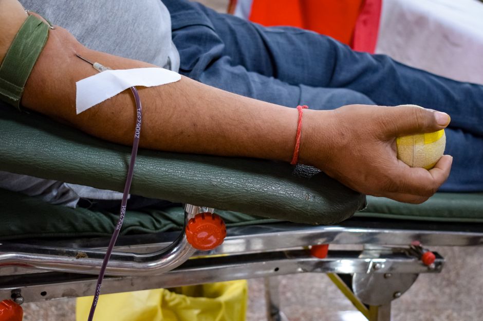
Photo by Rahul Sapra on Pexels
Blood doesn't just flow in one big loop; it travels through two main circuits:
- Pulmonary Circuit: This loop carries deoxygenated blood from the right side of your heart to your lungs to pick up oxygen and release carbon dioxide. The newly oxygenated blood then returns to the left side of your heart.
- Systemic Circuit: This much larger loop carries oxygenated blood from the left side of your heart to all the tissues and organs of your body, delivering oxygen and nutrients, and picking up waste. Deoxygenated blood then returns to the right side of your heart to restart the cycle.
This division ensures efficient oxygen delivery and waste removal.
graph TD
    A["Right Atrium"] --> B["Right Ventricle"]
    B --> C["Pulmonary Artery (to Lungs)"]
    C --> D["Lungs (Oxygenation)"]
    D --> E["Pulmonary Veins (from Lungs)"]
    E --> F["Left Atrium"]
    F --> G["Left Ventricle"]
    G --> H["Aorta (to Body)"]
    H --> I["Body Tissues (Exchange)"]
    I --> J["Vena Cavae (from Body)"]
    J --> A
    subgraph Pulmonary Circuit
        C --> D
        D --> E
    end
    subgraph Systemic Circuit
        H --> I
        I --> J
    end
    style A fill:#f9f,stroke:#333,stroke-width:2px
    style B fill:#f9f,stroke:#333,stroke-width:2px
    style F fill:#9ff,stroke:#333,stroke-width:2px
    style G fill:#9ff,stroke:#333,stroke-width:2px
3. Worked Example
Let's trace a single red blood cell's journey, starting from when it's just picked up oxygen in the lungs:
- The red blood cell (now oxygenated) travels from the lungs through the pulmonary veins.
- It enters the left atrium of the heart.
- From the left atrium, it moves into the left ventricle.
- The left ventricle contracts forcefully, ejecting the red blood cell into the aorta.
- The aorta branches into smaller arteries, carrying the red blood cell to a specific body tissue, let's say a muscle in your arm.
- In the muscle's capillaries, the red blood cell releases its oxygen to the muscle cells and picks up carbon dioxide.
- Now deoxygenated, the red blood cell moves into a venule, which merges into larger veins.
- It travels through progressively larger veins until it reaches one of the vena cavae.
- The vena cavae deliver the deoxygenated red blood cell to the right atrium of the heart.
- From the right atrium, it passes into the right ventricle.
- The right ventricle contracts, pumping the red blood cell into the pulmonary artery, which takes it back to the lungs to pick up oxygen again. The cycle repeats!
4. Key Takeaways
- The cardiovascular system is vital for transporting oxygen, nutrients, hormones, and waste products.
- It consists of the heart (pump), blood vessels (pipes), and blood (fluid).
- The heart has four chambers: right atrium, right ventricle, left atrium, and left ventricle.
- Arteries carry blood away from the heart, veins carry blood towards the heart, and capillaries are where exchange occurs.
- The pulmonary circuit moves blood between the heart and lungs, while the systemic circuit moves blood between the heart and the rest of the body.
- The right side of the heart handles deoxygenated blood, and the left side handles oxygenated blood.
Common mistakes to avoid:
* Confusing arteries with veins – remember Arteries go Away from the heart.
* Forgetting the heart's two-sided function (separate circuits for oxygenated/deoxygenated blood).
* Underestimating the role of capillaries as the actual sites of exchange.
* Mixing up the direction of blood flow in the two circuits (e.g., thinking pulmonary arteries carry oxygenated blood).
5. Now Try It
Draw a simplified diagram of the heart and its connections to the pulmonary and systemic circuits. Label the four chambers of the heart, the aorta, pulmonary artery, pulmonary veins, and vena cavae. Indicate with blue and red lines (or words) which vessels carry deoxygenated and oxygenated blood, respectively.
Success looks like: A clear, labeled diagram showing the two separate circuits, correct chamber names, and accurate representation of oxygenated vs. deoxygenated blood pathways.
Frequently asked about Introduction to the Cardiovascular System and Basic Anatomy
Study this next
Get the full Cardiovascular physiological curriculum
Clone the complete plan to your dashboard for unlimited AI-generated notes, practice quizzes, and a personalised revision schedule.
Save this course free