Anatomy of the Respiratory Tract
From the bio respiratory sys .in man curriculum
TL;DR
Your respiratory tract is like a branching tree, starting big and getting smaller, designed to efficiently get oxygen into your blood and remove carbon dioxide. It's divided into upper and lower sections, each with specific jobs for air conditioning and gas exchange. Understanding its structure helps you see how you breathe and stay healthy.
1. The Mental Model
Think of your respiratory system as a specialized pipeline for air. It's built to warm, humidify, and filter the air you breathe before it reaches tiny sacs in your lungs where oxygen jumps into your bloodstream.
2. The Core Material
Your respiratory tract is essentially the path air takes from the outside world into your lungs. It's typically divided into two main sections: the upper respiratory tract and the lower respiratory tract.
The Upper Respiratory Tract

Photo by Pavel Danilyuk on Pexels
This section includes your nose, nasal cavity, pharynx (throat), and larynx (voice box). Its main job is to prepare the air you breathe for your lungs.
- Nose and Nasal Cavity: Air first enters here. Hairs (cilia) and mucus trap dust and particles. A rich blood supply warms the air, and moisture from the mucus humidifies it.
- Pharynx (Throat): This is a shared pathway for both air and food. It's divided into three parts:
- Nasopharynx: Behind the nasal cavity, only for air.
- Oropharynx: Behind the oral cavity, for both air and food.
- Laryngopharynx: The lowest part, also for both air and food, leading to the esophagus (food pipe) and larynx.
- Larynx (Voice Box): This structure contains your vocal cords and is crucial for speech. It also acts as a gatekeeper, preventing food from entering your trachea (windpipe) during swallowing thanks to a flap called the epiglottis.
The Lower Respiratory Tract
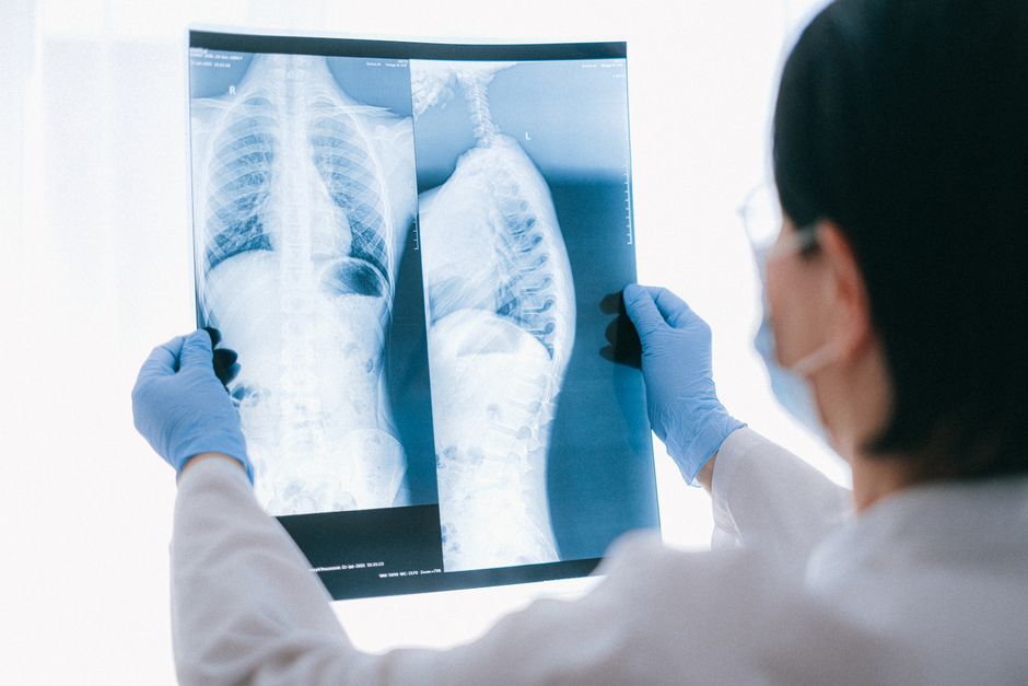
Photo by Anna Shvets on Pexels
This section starts from your trachea and goes into your lungs, where the real work of gas exchange happens.
- Trachea (Windpipe): This is a tube made of C-shaped cartilage rings that keep it open. It extends from the larynx down into your chest. Its inner lining has cilia and mucus to continue filtering air.
- Bronchi: The trachea splits into two main bronchi (one for each lung), which then branch into smaller and smaller tubes called bronchioles, much like the branches of a tree.
- Lungs: These are the primary organs of respiration, protected by your rib cage. Each lung is enclosed in a double-layered membrane called the pleura.
- Bronchioles: These are the smallest air passages, lacking cartilage. They lead to tiny air sacs.
- Alveoli: These are microscopic air sacs at the very end of the bronchioles. This is where oxygen diffuses into your bloodstream, and carbon dioxide diffuses out. The walls of the alveoli are incredibly thin, allowing for efficient gas exchange. They are surrounded by a network of capillaries (tiny blood vessels).
graph TD
    A["Air (Outside)"] --> B["Nose/Nasal Cavity"]
    B --> C["Pharynx"]
    C --> D["Larynx (Voice Box)"]
    D --> E["Trachea (Windpipe)"]
    E --> F["Main Bronchi (L/R)"]
    F --> G["Bronchioles"]
    G --> H["Alveoli (Gas Exchange)"]
    H --> I["Bloodstream (O2 in, CO2 out)"]
3. Worked Example
Let's trace a breath of air. When you inhale, air first enters your nose, where hairs filter out large particles, and tiny blood vessels warm and humidify it. It then passes through your pharynx, a shared pathway, and then into your larynx, past your vocal cords. From there, it descends into your trachea, kept open by its cartilage rings. The trachea branches into two main bronchi, which further divide into smaller bronchioles. Finally, the air reaches the thousands of tiny alveoli in your lungs, where oxygen diffuses across their thin walls into the surrounding capillaries and into your bloodstream, while carbon dioxide moves out of the blood and into the alveoli to be exhaled.
4. Key Takeaways
- The respiratory tract is divided into upper (nose to larynx) and lower (trachea to alveoli) sections.
- The upper tract cleans, warms, and humidifies incoming air.
- The lower tract includes the trachea, bronchi, bronchioles, and alveoli, facilitating gas exchange.
- The larynx protects the airway and enables speech with vocal cords.
- Alveoli are the primary sites where oxygen enters your blood and carbon dioxide leaves.
- Cartilage rings in the trachea and bronchi keep the airways open.
Common Mistakes to Avoid

Photo by KATRIN BOLOVTSOVA on Pexels
- Don't confuse the pharynx (throat, common to food and air) with the larynx (voice box, air only path).
- Remember the epiglottis is part of the larynx, not a separate organ.
- Don't forget that the bronchioles lack cartilage, unlike the trachea and larger bronchi.
- Overlooking the importance of mucus and cilia for air filtration throughout the tract.
5. Now Try It
Draw your own simplified diagram of the respiratory tract from memory, labeling at least eight major structures in the correct order that air would travel. Once done, describe in your own words the primary function of each labeled structure. You'll know you've succeeded if your diagram logically follows the path of air and your descriptions accurately reflect the roles of each part.
Frequently asked about Anatomy of the Respiratory Tract
Study this next
Get the full bio respiratory sys .in man curriculum
Clone the complete plan to your dashboard for unlimited AI-generated notes, practice quizzes, and a personalised revision schedule.
Save this course free