The Appendicular Skeleton: Pectoral and Pelvic Girdles
From the nursing curriculum
TL;DR
The appendicular skeleton consists of your limbs and the girdles that attach them to your axial skeleton. The pectoral girdle connects your upper limbs, providing flexibility, while the pelvic girdle connects your lower limbs, offering stability and weight bearing. Both girdles are crucial for movement and support throughout your body.
1. The Mental Model
Think of the appendicular skeleton as all the parts of your skeleton that append to your main body axis, letting you move around. The pectoral and pelvic girdles are like the strong, anchor points that connect your arms and legs to your trunk.
2. The Core Material
The appendicular skeleton includes the bones of your upper and lower limbs, and the girdles that attach them to the axial skeleton (your skull, vertebral column, and rib cage). We'll focus on these two crucial girdles.
The Pectoral Girdle (Shoulder Girdle)

Photo by Ekaterina Swiss on Pexels
The pectoral girdle attaches your upper limbs to the axial skeleton. It's designed for mobility and flexibility, allowing a wide range of motion for your arms.
It consists of two bones on each side:
1. Clavicle (Collarbone): An S-shaped bone that extends horizontally across the superior anterior thorax. It acts as a strut, holding the scapula and arm away from the trunk.
2. Scapula (Shoulder Blade): A flat, triangular bone located on the posterior aspect of the thorax. It articulates with the humerus (upper arm bone) at the glenoid cavity and with the clavicle.
Key Features of the Scapula:
* Glenoid Cavity: A shallow socket that articulates with the head of the humerus, forming the shoulder joint. This shallow design allows great mobility but also makes the joint prone to dislocation.
* Spine: A prominent ridge on the posterior surface that ends laterally in the acromion.
* Acromion: The lateral extension of the scapular spine that articulates with the clavicle.
* Coracoid Process: A hook-like projection anterior to the glenoid cavity, serving as an attachment point for muscles.
The Pelvic Girdle (Hip Girdle)
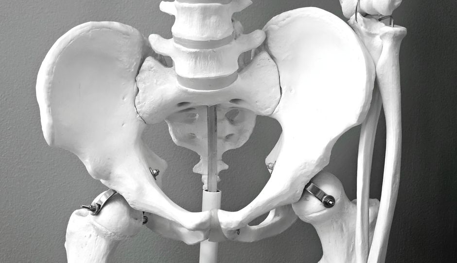
Photo by Magda Ehlers on Pexels
The pelvic girdle attaches your lower limbs to the axial skeleton. Unlike the pectoral girdle, it's built for strength, stability, and weight bearing. It also protects the organs of the pelvic cavity.
It's formed by two hip bones (also called coxal bones or os coxae) that articulate anteriorly at the pubic symphysis and posteriorly with the sacrum (part of your axial skeleton) at the sacroiliac joints. Each hip bone is a fusion of three separate bones during development:
1. Ilium: The largest and most superior part of the hip bone. You can feel its superior margin, the iliac crest, when you put your hands on your hips.
2. Ischium: The posterior and inferior part of the hip bone. When you sit down, you're sitting on your ischial tuberosities ("sit bones").
3. Pubis: The anterior and inferior part of the hip bone. The two pubic bones meet anteriorly to form the pubic symphysis.
Key Features of the Pelvic Girdle:
* Acetabulum: A deep, cup-shaped socket formed by the fusion of the ilium, ischium, and pubis. It articulates with the head of the femur (thigh bone), forming the hip joint. This deep socket provides excellent stability, reducing the risk of dislocation compared to the shoulder.
* Obturator Foramen: A large opening in the hip bone, partially closed by a membrane, that allows for the passage of nerves and blood vessels.
* Pelvic Brim (Pelvic Inlet): The boundary between the true (lesser) pelvis and the false (greater) pelvis. This is clinically important, especially in obstetrics.
* Greater Sciatic Notch: A large indentation on the posterior ilium, through which the sciatic nerve passes.
Differences Between Male and Female Pelvis
The pelvis has distinct structural differences between sexes, primarily related to childbirth in females.
graph TD
    A["Pelvic Girdle Function"] --> B["Pectoral Girdle (Upper Limb)"]
    A --> C["Pelvic Girdle (Lower Limb)"]

    B --> B1["Components: Clavicle, Scapula"]
    B --> B2["Primary Function: Mobility, Flexibility"]
    B --> B3["Joint: Shallow Glenoid Cavity (Shoulder)"]

    C --> C1["Components: Ilium, Ischium, Pubis (Fused as Hip Bone)"]
    C --> C2["Primary Function: Stability, Weight Bearing, Protection"]
    C --> C3["Joint: Deep Acetabulum (Hip)"]
    C --> C4["Articulation with Sacrum (Axial Skeleton)"]
    C --> C5["Sex Differences (e.g., Wider Female Pelvis)"]
3. Worked Example
Let's imagine a patient presenting with a dislocated shoulder versus a dislocated hip.
Scenario 1: Dislocated Shoulder
A young athlete falls directly onto their outstretched arm, resulting in a dislocated shoulder.
* Anatomy Involved: The head of the humerus has popped out of the glenoid cavity of the scapula.
* Why it happens: The pectoral girdle, particularly the glenoid cavity, is shallow. This anatomical design prioritizes a wide range of motion (flexion, extension, abduction, adduction, rotation) over stability. While strong ligaments and rotator cuff muscles provide support, a significant force can overcome them.
* Clinical Relevance: Shoulder dislocations are relatively common due to this design. Reduction (putting the bone back in place) is usually straightforward, but proper aftercare is needed to strengthen supporting structures.
Scenario 2: Dislocated Hip
A patient is involved in a severe car accident, and their knee strikes the dashboard, forcing their femur posteriorly, resulting in a dislocated hip.
* Anatomy Involved: The head of the femur has been forcefully dislodged from the acetabulum of the hip bone.
* Why it happens: The pelvic girdle, and specifically the acetabulum, is a very deep socket designed for immense stability and weight bearing. Dislocation requires significant force, often indicating severe trauma.
* Clinical Relevance: Hip dislocations are much less common than shoulder dislocations because of the inherent stability of the joint. When they occur, they are often associated with other serious injuries and require immediate medical attention, often under general anesthesia, due to the strong muscles and ligaments holding the joint.
This example highlights how the structural differences in the girdles (shallow vs. deep socket) directly correlate with their primary functions (mobility vs. stability) and influence the types of injuries seen.
4. Key Takeaways
- The appendicular skeleton includes the limbs and the girdles connecting them to the axial skeleton.
- The pectoral girdle (clavicle and scapula) provides a highly mobile attachment for the upper limbs.
- The shallow glenoid cavity of the scapula allows extensive shoulder movement but sacrifices stability.
- The pelvic girdle (fused ilium, ischium, pubis) forms a strong, stable base for the lower limbs.
- The deep acetabulum of the hip bone ensures strong articulation with the femur for weight bearing and stability.
- Male and female pelves have anatomical differences, primarily related to childbirth.
Common Mistakes to Avoid:
- Don't confuse the primary function of the pectoral girdle (mobility) with that of the pelvic girdle (stability).
- Remember the specific bones of each girdle (clavicle/scapula for pectoral; ilium/ischium/pubis for pelvic).
- Don't forget that the hip bone is actually three fused bones, not just one.
- Avoid thinking the sacrum is part of the pelvic girdle; it's part of the axial skeleton, but the hip bones articulate with it.
5. Now Try It
Take 15 minutes to draw a simple sketch of the anterior view of the pectoral girdle and the anterior view of the pelvic girdle. Label the major bones (clavicle, scapula, ilium, ischium, pubis) and key articulation points (glenoid cavity, acetabulum, pubic symphysis). What success looks like: You can identify and correctly label these structures, and briefly explain the main functional difference (mobility vs. stability) between the two girdles based on your drawings.
Frequently asked about The Appendicular Skeleton: Pectoral and Pelvic Girdles
Study this next
Get the full nursing curriculum
Clone the complete plan to your dashboard for unlimited AI-generated notes, practice quizzes, and a personalised revision schedule.
Save this course free