The Axial Skeleton: Skull and Vertebral Column
From the nursing curriculum
TL;DR
The axial skeleton forms the central axis of your body, protecting vital organs and providing support. It consists of the skull, vertebral column, and thoracic cage, which work together for protection, posture, and movement. Understanding these structures is key to grasping overall human anatomy and potential injuries.
1. The Mental Model
Think of the axial skeleton as your body's sturdy, protective core, like the main support beam of a house. It keeps you upright, shields your brain and spinal cord, and serves as an anchor for limbs, allowing you to move and function.
2. The Core Material
The axial skeleton includes the skull, vertebral column, and the thoracic cage (ribs and sternum). We'll focus on the skull and vertebral column here.
The Skull: Your Brain's Bony Helmet
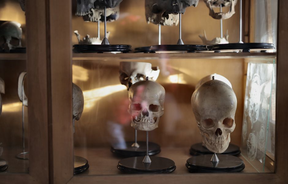
Photo by Andrea Piacquadio on Pexels
The skull is a complex structure made of 22 bones, divided into two main parts: the cranium and the facial bones.
- Cranium (8 bones): These bones encase and protect the brain. They include the frontal, parietal (2), temporal (2), occipital, sphenoid, and ethmoid bones. They're mostly flat bones joined by immovable joints called sutures.
- Facial Bones (14 bones): These form the face, provide attachment for facial muscles, and house sensory organs (eyes, nose, mouth). Key bones include the maxillae (upper jaw), zygomatic bones (cheekbones), nasal bones, and the mandible (lower jaw, the only movable skull bone).
Key Features of the Skull:
* Foramina: Openings for nerves and blood vessels (e.g., foramen magnum for the spinal cord).
* Sinuses: Air-filled cavities within some cranial and facial bones that lighten the skull and resonate sound.
* Sutures: Immovable joints that fuse skull bones together (e.g., coronal, sagittal, lambdoid).
The Vertebral Column: Your Body's Flexible Support Rod

Photo by cottonbro studio on Pexels
Also known as the spine or backbone, the vertebral column extends from the skull to the pelvis. It consists of 33 individual bones called vertebrae (though some fuse in adulthood), separated by intervertebral discs. It provides support, allows for movement, and protects the delicate spinal cord.
The vertebral column is divided into five main regions:
-
Cervical Vertebrae (C1-C7):
- 7 vertebrae in the neck.
- Smallest and most delicate.
- C1 (Atlas) supports the skull, allowing for "yes" nodding.
- C2 (Axis) has the dens (odontoid process), allowing for "no" head rotation.
- Have transverse foramina for vertebral arteries.
-
Thoracic Vertebrae (T1-T12):
- 12 vertebrae in the upper back.
- Articulate with the ribs, providing attachment points for the thoracic cage.
- Have longer spinous processes that point inferiorly.
-
Lumbar Vertebrae (L1-L5):
- 5 vertebrae in the lower back.
- Largest and strongest vertebrae, bearing most of the body's weight.
- Have short, blunt spinous processes.
-
Sacrum (5 fused vertebrae):
- A triangular bone formed by the fusion of 5 sacral vertebrae.
- Articulates with the ilium of the pelvic girdle.
-
Coccyx (3-5 fused vertebrae):
- Your "tailbone," formed by the fusion of 3-5 tiny vertebrae.
- A remnant of a tail, serves as an attachment point for some muscles.
Curves of the Vertebral Column:
The spine has four natural curves, which increase its strength, flexibility, and ability to absorb shock:
* Cervical and Lumbar curves: Lordotic (curve anteriorly).
* Thoracic and Sacral curves: Kyphotic (curve posteriorly).
Intervertebral Discs:
These are fibrocartilaginous pads between vertebrae, acting as shock absorbers and allowing for spinal flexibility. Each disc has two parts:
* Nucleus Pulposus: Inner, gel-like core.
* Anulus Fibrosus: Outer ring of tough fibrocartilage.
Here's how the different regions of the vertebral column are structured:
graph TD
    A["Vertebral Column"] --> B["Cervical (C1-C7)"]
    A --> C["Thoracic (T1-T12)"]
    A --> D["Lumbar (L1-L5)"]
    A --> E["Sacrum (5 fused)"]
    A --> F["Coccyx (3-5 fused)"]

    B --> B1["Atlas (C1)"]
    B --> B2["Axis (C2)"]
    C --> C1["Rib Articulation"]
    D --> D1["Weight Bearing"]
    E --> E1["Pelvic Girdle"]
    F --> F1["Muscle Attachment"]
3. Worked Example
Imagine a patient comes into the ER after a fall, complaining of severe neck pain and difficulty moving their head.
- Assessment: The doctor orders X-rays and potentially a CT scan of the cervical spine.
- Findings: The imaging reveals a fracture of the dens of the Axis (C2) vertebra.
- Implications: A fracture of the dens is serious because it directly affects the stability of the atlas and the ability to rotate the head. It's crucial for protecting the spinal cord.
- Nursing Considerations:
- Immobilization: The patient will require immediate immobilization with a cervical collar or halo vest to prevent further injury to the spinal cord.
- Pain Management: Administer prescribed analgesics and assess pain levels regularly.
- Neurological Monitoring: Closely monitor for any changes in sensation, motor function, or reflexes in the extremities, indicating potential spinal cord compression.
- Patient Education: Educate the patient and family about the injury, the need for immobilization, and how to safely move and care for themselves with the device.
Understanding the specific anatomy of C2 (Axis) and its role in head movement and spinal cord protection is critical for proper assessment and care in this scenario.
4. Key Takeaways
- The axial skeleton provides the central framework, protecting your brain and spinal cord, and supporting the body.
- The skull's cranium shields the brain, while facial bones form the face and house sensory organs.
- Skull bones are joined by sutures, except for the movable mandible (jaw).
- The vertebral column, made of cervical, thoracic, lumbar, sacral, and coccygeal regions, offers support and flexibility.
- Intervertebral discs cushion the vertebrae and allow for spinal movement.
- The natural curves of the spine (lordotic and kyphotic) enhance its strength and shock-absorbing capacity.
Common Mistakes to Avoid:
- Don't confuse the cranium with the entire skull; the skull also includes facial bones.
- Misunderstanding that C1 (Atlas) allows nodding, and C2 (Axis) with its dens allows rotation.
- Forgetting the protective role of the vertebral column for the spinal cord, especially in injury situations.
- Overlooking the importance of the intervertebral discs for flexibility and shock absorption.
5. Now Try It
Take 15 minutes to draw a simplified sketch of the human vertebral column. Label the five main regions (cervical, thoracic, lumbar, sacrum, coccyx) and indicate where the natural curves are. For an extra challenge, try to roughly sketch and label the Atlas and Axis vertebrae. Success looks like accurately identifying each region and their respective curvatures.
Frequently asked about The Axial Skeleton: Skull and Vertebral Column
Study this next
Get the full nursing curriculum
Clone the complete plan to your dashboard for unlimited AI-generated notes, practice quizzes, and a personalised revision schedule.
Save this course free