The Five P's of Labor: Passage
From the Maternal curriculum
TL;DR
"Passage" in labor refers to the birth canal, which includes the bony pelvis and soft tissues. The size and shape of your client's pelvis, along with the ability of soft tissues to stretch, directly impact how the baby descends during labor. Understanding pelvic types and tissue flexibility helps predict potential challenges and guide interventions.
1. The Mental Model
Think of the birth canal as a tunnel. For a smooth delivery, the baby (passenger) needs to fit through this tunnel (passage), which can change shape slightly, and the surrounding tissues need to be flexible enough to allow passage.
2. The Core Material
"Passage" is one of the crucial "P's" because it represents the route the baby must take from the uterus to the outside world. It involves two main components: the bony pelvis and the soft tissues.
The Bony Pelvis
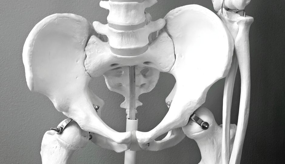
Photo by Magda Ehlers on Pexels
The bony pelvis is the primary determinant of the passage. It's a rigid structure, but its individual bones (ilium, ischium, pubis, sacrum, coccyx) are connected by ligaments that allow for slight movement, especially as labor progresses.
There are four main pelvic types, and while most people have a mixed type, one usually predominates:
- Gynecoid: This is considered the "classic female pelvis" and is the most favorable for vaginal birth. It's rounded and wide.
- Anthropoid: Oval-shaped with a wider front-to-back diameter, but narrower side-to-side. Can also allow for vaginal birth, often with a posterior presentation.
- Android: Heart-shaped, like a male pelvis, and more narrow overall. This type often presents challenges for vaginal birth due to reduced space.
- Platypelloid: Flat and wide side-to-side, but narrow front-to-back. This is the least common type and often makes vaginal birth difficult.
Pelvic Planes
The baby descends through three main planes within the pelvis:
- Inlet (Pelvic Brim): The top opening of the pelvis. The widest diameter here is usually transverse (side-to-side).
- Midpelvis (Pelvic Cavity): The narrowest part of the pelvis, typically at the ischial spines. This is a critical point for fetal rotation.
- Outlet: The bottom opening of the pelvis. The widest diameter here is usually anteroposterior (front-to-back).
The baby must navigate these varying diameters, rotating as needed to fit through the tightest spots.
The Soft Tissues

Photo by Magda Ehlers on Pexels
Beyond the bony pelvis, the soft tissues also form part of the passage. These include:
- Lower uterine segment: The bottom part of the uterus that stretches and thins during labor.
- Cervix: The opening of the uterus, which must efface (thin) and dilate (open) to 10 cm.
- Vagina: The muscular canal that connects the uterus to the outside.
- Perineum: The area between the vagina and rectum, which needs to stretch and sometimes tear or be episiotomized to allow passage.
The flexibility and ability of these tissues to stretch are crucial. Scar tissue from previous surgeries or injuries can sometimes make passage more difficult.
Here's how the baby navigates the passage:
graph TD
    A["Fetal Head Engages (Pelvic Inlet)"] --> B["Descent"]
    B --> C["Flexion"]
    C --> D["Internal Rotation (to fit Midpelvis)"]
    D --> E["Extension (under Symphysis Pubis)"]
    E --> F["Restitution (External Rotation)"]
    F --> G["Expulsion (Shoulders & Body)"]
3. Worked Example
Imagine your client, Sarah, is in active labor. During her prenatal visits, a pelvic exam suggested she has a generally gynecoid pelvis, which is favorable. As she progresses, you notice the fetal head is at +2 station, meaning it's past the ischial spines and well into the midpelvis. However, during pushing, progress seems to slow.
Upon reassessment, you observe that the baby's head is still quite high for the pushing phase, and Sarah reports significant pressure in her sacrum. This could indicate a subtle challenge with the pelvic outlet, perhaps a slightly less roomy anteroposterior diameter than ideal, even within a generally gynecoid type.
Your interventions might include:
- Changing maternal positions: Encouraging positions like hands-and-knees or squatting can help open the pelvic outlet.
- Applying counterpressure: To relieve sacral pain and potentially encourage slight sacral movement.
- Assessing for soft tissue edema: Excessive swelling of the perineum could impede progress.
By focusing on the "Passage" (in this case, the pelvic outlet and potential soft tissue issues), you can proactively help Sarah achieve a vaginal birth by optimizing the available space.
4. Key Takeaways
- The birth canal (passage) consists of the rigid bony pelvis and flexible soft tissues.
- Pelvic shape (gynecoid, anthropoid, android, platypelloid) significantly impacts labor progress.
- The baby must navigate different diameters within the pelvic inlet, midpelvis, and outlet.
- Soft tissues like the cervix, vagina, and perineum must stretch and open for delivery.
- Previous injuries or scar tissue can affect soft tissue flexibility and passage.
- Understanding the passage helps you anticipate challenges and guide interventions like maternal positioning.
5. Now Try It
For your next client in labor, actively observe and document your assessment of "Passage." Consider their reported pain, any pelvic exam findings (station, effacement, dilation), and how their chosen labor positions might be affecting the space available for the baby. After your shift, reflect on how your understanding of "Passage" influenced your care decisions. Success looks like you identifying one specific aspect of "Passage" that was relevant to your client's labor progress and how you responded to it.
Frequently asked about The Five P's of Labor: Passage
Study this next
Get the full Maternal curriculum
Clone the complete plan to your dashboard for unlimited AI-generated notes, practice quizzes, and a personalised revision schedule.
Save this course free