Introduction to the Brachial Plexus and Cervical Anatomy
From the Brachial plexus curriculum
TL;DR
The brachial plexus is a network of nerves originating from the lower cervical and upper thoracic spinal cord, responsible for innervating your entire upper limb. It forms a complex structure that's essential for movement and sensation in your arm and hand. Understanding its anatomical relationship with the cervical spine is key to grasping its function and potential clinical issues.
1. The Mental Model
Think of the brachial plexus as your arm's "power cable" that branches out into many smaller wires. This main cable originates from your neck, and any issue along its path, from the spinal cord to the arm, can impact how your arm and hand work.
2. The Core Material
The brachial plexus is an intricate network formed by the anterior rami (ventral rami) of spinal nerves C5, C6, C7, C8, and T1. Sometimes, contributions from C4 and T2 might also be present, which are called "prefixed" or "postfixed" plexuses, respectively. This network undergoes a series of divisions and recombinations as it travels from the neck through the axilla (armpit) to supply the upper limb.
Let's break down its structure into five main sections, often remembered by the mnemonic "Randy Travis Drinks Cold Beer":
- Roots: These are the anterior rami of the spinal nerves (C5, C6, C7, C8, T1) as they emerge from the intervertebral foramina between the vertebrae.
- Trunks: The roots combine to form three trunks:
- Upper trunk: C5 + C6
- Middle trunk: C7 (continues directly)
- Lower trunk: C8 + T1
- Divisions: Each trunk then splits into an anterior and a posterior division. There are six divisions in total (3 anterior, 3 posterior). These divisions are crucial for determining which muscles will be supplied. Anterior divisions generally supply flexor compartments, while posterior divisions supply extensor compartments.
- Cords: The divisions recombine to form three cords, named for their relationship to the axillary artery in the axilla:
- Lateral cord: Formed by the anterior divisions of the upper and middle trunks.
- Posterior cord: Formed by all three posterior divisions (from upper, middle, and lower trunks).
- Medial cord: Formed by the anterior division of the lower trunk.
- Branches: The cords give rise to the major peripheral nerves of the upper limb.
Cervical Anatomy Relationship
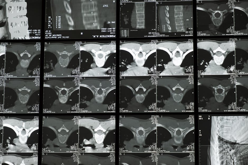
Photo by cottonbro studio on Pexels
The roots of the brachial plexus emerge from the cervical spine. Specifically:
* The C5 nerve root exits above the C5 vertebra.
* The C6 nerve root exits above the C6 vertebra.
* The C7 nerve root exits above the C7 vertebra.
* The C8 nerve root exits below the C7 vertebra (there are 7 cervical vertebrae but 8 cervical nerve roots).
* The T1 nerve root exits below the T1 vertebra.
These nerve roots pass between the anterior and middle scalene muscles in the neck, a common site for compression. They then pass over the first rib, beneath the clavicle, and into the axilla. This close anatomical relationship means that conditions affecting the cervical spine (like disc herniations, cervical ribs, or muscle spasms) can directly impact the brachial plexus.
graph TD
    subgraph Roots
        C5["C5"] --> UT
        C6["C6"] --> UT
        C7["C7"] --> MT
        C8["C8"] --> LT
        T1["T1"] --> LT
    end

    subgraph Trunks
        UT["Upper Trunk"] --> ADUT["Anterior Division (Upper)"]
        UT --> PDUT["Posterior Division (Upper)"]
        MT["Middle Trunk"] --> ADMT["Anterior Division (Middle)"]
        MT --> PDMT["Posterior Division (Middle)"]
        LT["Lower Trunk"] --> ADLT["Anterior Division (Lower)"]
        LT --> PDLT["Posterior Division (Lower)"]
    end

    subgraph Divisions
        ADUT --> LC["Lateral Cord"]
        ADMT --> LC
        ADLT --> MC["Medial Cord"]
        PDUT --> PC["Posterior Cord"]
        PDMT --> PC
        PDLT --> PC
    end

    subgraph Cords
        LC --> MMNA["Musculocutaneous Nerve"]
        LC --> LRMNA["Lateral Root of Median Nerve"]
        MC --> UNa["Ulnar Nerve"]
        MC --> MRMNA["Medial Root of Median Nerve"]
        MRMNA --> MNa["Median Nerve"]
        LRMNA --> MNa
        PC --> AxNa["Axillary Nerve"]
        PC --> RadNa["Radial Nerve"]
    end
3. Worked Example
Imagine a patient presents with weakness in their deltoid muscle (shoulder abduction) and numbness over the lateral aspect of their forearm.
Looking at our diagram:
1. Deltoid weakness (shoulder abduction): The deltoid muscle is innervated by the axillary nerve. The axillary nerve originates from the posterior cord. The posterior cord is formed by the posterior divisions of the upper, middle, and lower trunks.
2. Numbness over the lateral forearm: This area is typically supplied by the musculocutaneous nerve. The musculocutaneous nerve originates from the lateral cord. The lateral cord is formed by the anterior divisions of the upper and middle trunks.
3. Connecting the dots: Both the axillary nerve and musculocutaneous nerve receive contributions from the upper trunk (C5, C6). If there's an issue affecting the C5 or C6 nerve roots, or the upper trunk itself, both these symptoms could manifest. This could point to a lesion high up in the plexus, such as a disc herniation affecting C5/C6 nerve roots or an injury to the upper trunk.
4. Key Takeaways
- The brachial plexus is formed by spinal nerves C5-T1, supplying the entire upper limb.
- Its structure progresses from Roots, Trunks, Divisions, Cords, to Branches (Randy Travis Drinks Cold Beer).
- The roots emerge from the cervical spine, with C8 exiting below C7.
- Divisions determine muscle compartment supply: anterior for flexors, posterior for extensors.
- Cords are named for their relationship to the axillary artery and give rise to major peripheral nerves.
- Cervical spine pathology (like disc herniation or scalene muscle compression) can directly impact brachial plexus function.
Common Mistakes to Avoid:
* Confusing the order of the brachial plexus components (R, T, D, C, B).
* Forgetting that C8 exits below C7, which is different from other cervical roots.
* Not understanding that a lesion high up (e.g., at the root or trunk level) can cause widespread deficits across multiple terminal nerves.
* Mixing up which divisions form which cords (e.g., posterior cord is all posterior divisions).
5. Now Try It
Take an empty piece of paper and draw the entire brachial plexus from memory, including the roots, trunks, divisions, cords, and the major terminal branches (musculocutaneous, axillary, radial, median, ulnar). Label the spinal nerve contributions to each trunk. Then, try to trace the path of the median nerve back to its roots. Success looks like accurately drawing and labeling the entire plexus without referring to notes, and correctly identifying the roots contributing to the median nerve.
Frequently asked about Introduction to the Brachial Plexus and Cervical Anatomy
Study this next
Get the full Brachial plexus curriculum
Clone the complete plan to your dashboard for unlimited AI-generated notes, practice quizzes, and a personalised revision schedule.
Save this course free