Introduction to Radiography and Radiation Physics
From the Radiography curriculum
TL;DR
Radiography uses X-rays, a type of electromagnetic radiation, to create images of the inside of the body. Understanding how X-rays are produced and how they interact with matter is fundamental to producing high-quality diagnostic images safely. Radiation physics explains these interactions, ensuring you can protect yourself and your patients while getting the best possible images.
1. The Mental Model
Think of radiography as using a special kind of light (X-rays) that can pass through your body. Different parts of your body block this light differently, creating a shadow image on a detector. Radiation physics is the science behind how this special light is made and how it interacts with the stuff it hits.
2. The Core Material
What is Radiography?

Photo by Gustavo Fring on Pexels
Radiography is the art and science of creating images of the internal structures of the body using X-rays. These images, called radiographs, help doctors diagnose and monitor various medical conditions. Your role as a radiographer is to produce these images safely and effectively.
What are X-rays?

Photo by cottonbro studio on Pexels
X-rays are a form of electromagnetic radiation, just like visible light, radio waves, or microwaves. The key difference is their much shorter wavelength and higher energy. This high energy allows them to penetrate materials that visible light cannot, like human tissue.
How X-rays are Produced
X-rays are produced in an X-ray tube. Here's a simplified breakdown:
1. Cathode (Negative Electrode): A filament (like in a light bulb) is heated, causing electrons to boil off (thermionic emission).
2. Anode (Positive Electrode): A high voltage accelerates these electrons towards a metal target (usually tungsten).
3. Collision: When the fast-moving electrons hit the anode target, their kinetic energy is converted into X-rays (about 1%) and heat (about 99%).
4. X-ray Emission: The X-rays then exit the tube through a window.
Radiation Physics: Interaction with Matter
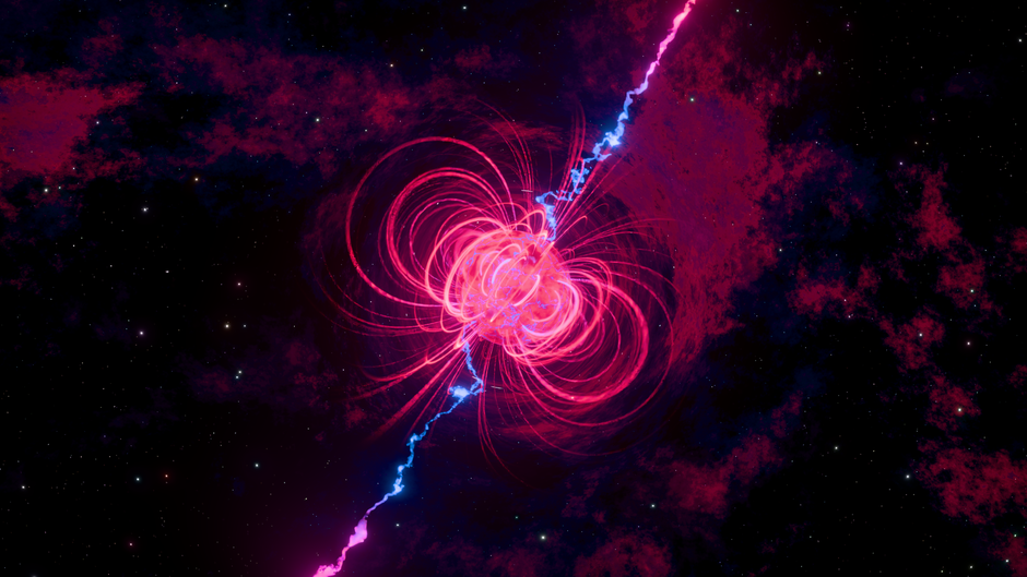
Photo by Nicola Narracci on Pexels
When X-rays pass through the body, they interact with tissues in several ways. These interactions are what create the image:
- Absorption (Photoelectric Effect): X-ray photons are completely absorbed by atoms in the tissue. This happens more in dense materials like bone.
- Scattering (Compton Effect): X-ray photons lose some energy and change direction. This contributes to image "fog" and increases patient dose.
- Transmission: X-ray photons pass through the tissue without interacting. This happens more in less dense materials like air or soft tissue.
The difference in absorption and transmission between various tissues creates the contrast on a radiograph. Bones appear white because they absorb more X-rays, while air-filled lungs appear black because they transmit most X-rays.
graph TD
    A["Electrons from Cathode"] --> B["Accelerated by High Voltage"]
    B --> C["Strike Anode Target (Tungsten)"]
    C --> D{"Energy Conversion"}
    D --> E["~1% X-rays Produced"]
    D --> F["~99% Heat Produced"]
    E --> G["X-rays Exit Tube"]
    G --> H["Interact with Patient Tissue"]
    H --> I{"Interaction Type"}
    I --> J["Absorption (e.g., Bone)"]
    I --> K["Scattering (Image Fog)"]
    I --> L["Transmission (e.g., Air)"]
    J --> M["Less X-rays Reach Detector (White Area)"]
    K --> M
    L --> N["More X-rays Reach Detector (Black Area)"]
Radiation Safety Principles (ALARA)

Photo by Jo McNamara on Pexels
Because X-rays can cause biological damage, radiation safety is paramount. The guiding principle is ALARA: As Low As Reasonably Achievable. This means always striving to minimize radiation exposure to patients, yourself, and others.
Key ALARA principles:
* Time: Reduce the duration of exposure.
* Distance: Increase the distance from the radiation source.
* Shielding: Use protective barriers (e.g., lead aprons, lead walls).
3. Worked Example
Imagine you're taking a chest X-ray.
1. You position the patient correctly between the X-ray tube and the image receptor.
2. When you activate the X-ray machine, electrons are fired from the cathode to the tungsten anode, generating X-rays.
3. These X-rays travel through the patient's chest.
* The ribs and spine (bone) absorb a lot of the X-rays (photoelectric effect), so fewer X-rays reach the detector in those areas.
* The lungs (mostly air) transmit most of the X-rays, so many X-rays reach the detector.
* The heart and diaphragm (soft tissue) absorb some X-rays, but less than bone and more than air.
4. The detector records the varying amounts of X-rays that pass through. Where few X-rays hit (bone), the image appears white. Where many X-rays hit (lungs), the image appears black. This difference creates the diagnostic image.
4. Key Takeaways
- Radiography uses high-energy X-rays to visualize internal body structures.
- X-rays are produced when fast-moving electrons hit a metal target in an X-ray tube.
- X-rays interact with matter through absorption, scattering, and transmission, which create the image contrast.
- Dense tissues like bone absorb more X-rays and appear white; less dense tissues like air transmit more X-rays and appear black.
- The ALARA principle (As Low As Reasonably Achievable) guides all radiation safety practices to minimize exposure.
- Understanding radiation physics is crucial for producing diagnostic images safely and effectively.
Common Mistakes to Avoid:
- Forgetting to properly shield the patient (e.g., using a lead apron for gonadal shielding).
- Not understanding the basic principles of X-ray production, which can lead to poor image quality.
- Overlooking the importance of distance and time in reducing your own radiation exposure.
- Assuming all radiation is bad; therapeutic uses exist, but diagnostic uses require careful management.
5. Now Try It
Spend 15 minutes researching the different types of X-ray tubes (e.g., stationary anode vs. rotating anode). For each type, briefly describe its main advantage and disadvantage, and consider in what common clinical situations each might be preferred. Success looks like being able to explain why a rotating anode tube is generally used for procedures requiring higher X-ray output compared to a stationary anode tube.
Frequently asked about Introduction to Radiography and Radiation Physics
Study this next
Get the full Radiography curriculum
Clone the complete plan to your dashboard for unlimited AI-generated notes, practice quizzes, and a personalised revision schedule.
Save this course free