Tissues: The Fabric of the Body
From the BIOL-2201-153:Human Anatomy & Physiology I • 39409.202630 curriculum
TL;DR
Your body is built from four basic tissue types: epithelial, connective, muscle, and nervous tissue, each with specialized functions. These tissues work together to form organs and systems, allowing your body to function. Understanding these tissue types is fundamental to grasping human anatomy and physiology.
1. The Mental Model
Think of tissues as specialized building blocks. Just as bricks, wood, and glass are used for different parts of a house, your body uses specific tissues to perform distinct jobs, from covering surfaces to enabling movement.
2. The Core Material
Your body is organized in a hierarchical way, starting from chemical levels up to the entire organism. Tissues are the next level up from cells, defined as groups of similar cells that perform a common function. There are four primary tissue types, each with unique characteristics and roles:
2.1 Epithelial Tissue (Epithelium)
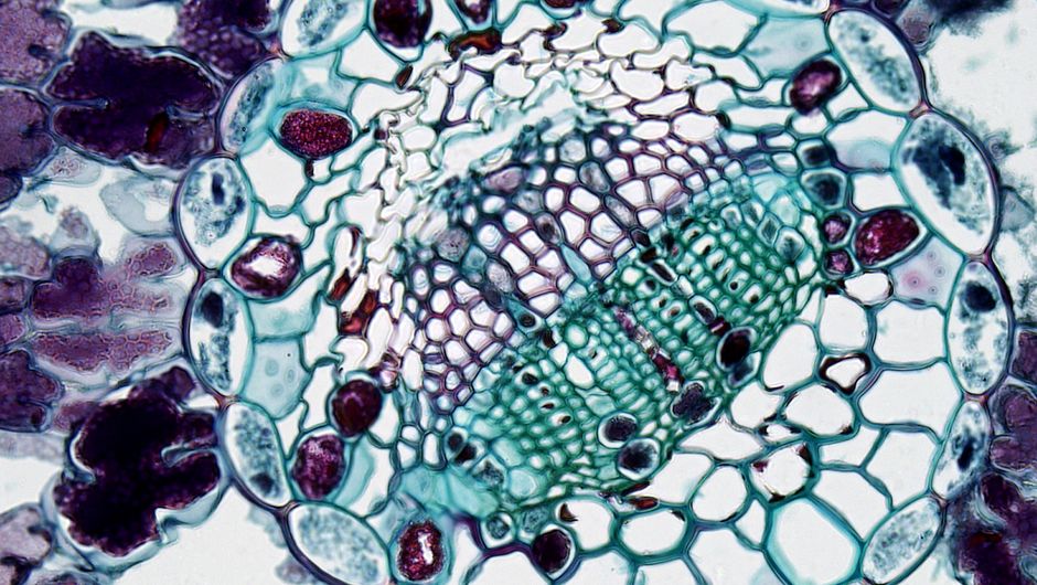
Photo by Fayette Reynolds M.S. on Pexels
This tissue forms coverings, linings, and glandular tissue throughout your body. Its main functions include protection, absorption, filtration, and secretion. Epithelial cells are tightly packed together, often forming sheets, and have a free surface (apical surface) exposed to the exterior or to a cavity, and an attached surface (basal surface). They are avascular (no direct blood supply) and rely on diffusion from underlying connective tissue for nutrients.
Epithelial tissue is classified by two main criteria:
* Number of cell layers:
* Simple: One layer of cells, involved in absorption, secretion, and filtration.
* Stratified: More than one layer of cells, primarily protective.
* Shape of cells:
* Squamous: Flat, scale-like.
* Cuboidal: Cube-shaped.
* Columnar: Column-shaped.
Examples include the epidermis of the skin (stratified squamous) and the lining of the digestive tract (simple columnar).
2.2 Connective Tissue

Photo by Fayette Reynolds M.S. on Pexels
This is the most abundant and widely distributed tissue type in your body. It primarily supports, protects, binds together other tissues, and fills spaces. Connective tissue has a unique structure: it's composed of cells (like fibroblasts, chondrocytes, osteocytes, adipocytes) widely scattered within an abundant extracellular matrix. This matrix consists of ground substance (fluid, gel-like, or solid material) and fibers (collagen, elastic, reticular). The type and amount of fibers and ground substance determine the tissue's function.
Major types of connective tissue include:
* Loose connective tissue: Areolar, adipose (fat), reticular.
* Dense connective tissue: Dense regular (tendons, ligaments), dense irregular (dermis), elastic.
* Cartilage: Hyaline, elastic, fibrocartilage.
* Bone: Compact, spongy.
* Blood: A liquid connective tissue.
2.3 Muscle Tissue

Photo by https://kaboompics.com/ on Pexels
Specialized for contraction, muscle tissue is responsible for body movement, moving substances through internal organs, and maintaining posture. There are three types:
* Skeletal muscle: Voluntary, striated (striped appearance), attached to bones.
* Cardiac muscle: Involuntary, striated, found only in the heart.
* Smooth muscle: Involuntary, non-striated, found in walls of internal organs (e.g., digestive tract, blood vessels).
2.4 Nervous Tissue
Photo by Fayette Reynolds M.S. on Pexels
Nervous tissue forms the brain, spinal cord, and nerves. Its primary function is to communicate by transmitting electrical signals (nerve impulses) rapidly throughout the body. It consists of two main cell types:
* Neurons: The excitable cells that transmit signals.
* Neuroglia (glial cells): Support, insulate, and protect neurons.
graph TD
    A["Organism"] --> B["Organ Systems"]
    B --> C["Organs"]
    C --> D["Tissues"]
    D --> E1["Epithelial Tissue"]
    D --> E2["Connective Tissue"]
    D --> E3["Muscle Tissue"]
    D --> E4["Nervous Tissue"]
    E1 --> F1a["Simple Squamous (lung air sacs)"]
    E1 --> F1b["Stratified Squamous (skin epidermis)"]
    E2 --> F2a["Loose Areolar (under epithelia)"]
    E2 --> F2b["Bone (skeleton)"]
    E3 --> F3a["Skeletal Muscle (biceps)"]
    E3 --> F3b["Cardiac Muscle (heart wall)"]
    E4 --> F4a["Neurons (brain)"]
    E4 --> F4b["Neuroglia (spinal cord)"]
3. Worked Example
Imagine you're looking at a cross-section of the small intestine. You'd see several tissue types working together. The innermost lining, facing the food, would be simple columnar epithelial tissue, specialized for absorbing nutrients. Beneath this, you'd find loose areolar connective tissue, providing support and housing blood vessels and nerves. Deeper still, layers of smooth muscle tissue would be present, contracting involuntarily to move food along. Finally, nervous tissue (part of the enteric nervous system) would be embedded within these layers, coordinating muscle contractions and gland secretions. Each tissue plays a vital, distinct role in the overall function of digestion.
4. Key Takeaways
- There are four main tissue types: epithelial, connective, muscle, and nervous.
- Epithelial tissue covers, lines, and forms glands, protecting and regulating passage of substances.
- Connective tissue supports, binds, protects, and fills space, characterized by an extracellular matrix.
- Muscle tissue is specialized for contraction, enabling movement.
- Nervous tissue transmits electrical signals for communication and control.
- Tissue classification helps predict function and location within the body.
- Different tissue types work together to form organs and perform complex functions.
Common mistakes to avoid:
- Confusing the functions of epithelial and connective tissue (e.g., thinking connective tissue forms coverings).
- Forgetting that blood is a type of connective tissue.
- Mixing up the voluntary/involuntary and striated/non-striated characteristics of muscle types.
- Not recognizing the importance of the extracellular matrix in connective tissue.
5. Now Try It
Choose any organ in your body (e.g., stomach, lung, skin). For that organ, try to identify at least two of the four basic tissue types that would be present, and describe their specific function within that organ.
What success looks like: You can correctly name at least two tissue types for your chosen organ and accurately describe their role, demonstrating an understanding of how tissue structure relates to organ function.
Frequently asked about Tissues: The Fabric of the Body
More from BIOL-2201-153:Human Anatomy & Physiology I • 39409.202630
Get the full BIOL-2201-153:Human Anatomy & Physiology I • 39409.202630 curriculum
Clone the complete plan to your dashboard for unlimited AI-generated notes, practice quizzes, and a personalised revision schedule.
Save this course free