The Human Heart
From the FSC Biology curriculum
TL;DR
The human heart is a muscular pump divided into four chambers that circulates blood throughout your body. It works in two main circuits: the pulmonary circuit for oxygenation and the systemic circuit for delivery to tissues. Understanding its structure and function is key to grasping how your body maintains life.
1. The Mental Model
Think of your heart as a tireless, two-sided pump. One side sends "used" blood to the lungs to pick up oxygen, while the other side sends fresh, oxygenated blood to every other part of your body. It's constantly working to keep everything moving.
2. The Core Material
Your heart is a fist-sized organ located slightly to the left of the center of your chest, protected by your rib cage. It's made of cardiac muscle, a special type of muscle tissue that can contract rhythmically and involuntarily.
The heart is divided into four chambers:
* Right Atrium (RA): Receives deoxygenated blood from the body.
* Right Ventricle (RV): Pumps deoxygenated blood to the lungs.
* Left Atrium (LA): Receives oxygenated blood from the lungs.
* Left Ventricle (LV): Pumps oxygenated blood to the rest of the body. This is the strongest chamber as it has to pump blood furthest.
These chambers are separated by valves that act like one-way doors, preventing blood from flowing backward.
* Tricuspid valve: Between RA and RV.
* Pulmonary valve: Between RV and pulmonary artery.
* Mitral (bicuspid) valve: Between LA and LV.
* Aortic valve: Between LV and aorta.
Blood Flow and Circuits

Photo by Landiva Weber on Pexels
Blood circulates through your body in two main pathways:
The Pulmonary Circuit
This circuit carries deoxygenated blood from the heart to the lungs and then returns oxygenated blood to the heart.
1. Deoxygenated blood from the body enters the right atrium.
2. It passes through the tricuspid valve into the right ventricle.
3. The right ventricle pumps it through the pulmonary valve into the pulmonary artery, which takes it to the lungs.
4. In the lungs, blood releases carbon dioxide and picks up oxygen.
5. Oxygenated blood returns to the left atrium via the pulmonary veins.
The Systemic Circuit
This circuit carries oxygenated blood from the heart to the rest of the body and returns deoxygenated blood to the heart.
1. Oxygenated blood from the lungs enters the left atrium.
2. It passes through the mitral valve into the left ventricle.
3. The left ventricle pumps it through the aortic valve into the aorta, the body's largest artery.
4. The aorta branches into smaller arteries, delivering oxygenated blood to all tissues and organs.
5. After oxygen is delivered and waste products (like CO2) are picked up, deoxygenated blood returns to the heart via veins, eventually collecting in the vena cavae (superior and inferior) which empty into the right atrium.
Here's a diagram illustrating the path of blood through the heart and the two circuits:
graph TD
    A["Body (Deoxygenated Blood)"] --> VSC["Vena Cavae (Superior/Inferior)"]
    VSC --> RA["Right Atrium"]
    RA --> TV["Tricuspid Valve"]
    TV --> RV["Right Ventricle"]
    RV --> PV["Pulmonary Valve"]
    PV --> PA["Pulmonary Artery"]
    PA --> L["Lungs (Gas Exchange)"]
    L --> PVN["Pulmonary Veins"]
    PVN --> LA["Left Atrium"]
    LA --> MV["Mitral Valve"]
    MV --> LV["Left Ventricle"]
    LV --> AV["Aortic Valve"]
    AV --> Aorta["Aorta"]
    Aorta --> B["Body (Oxygenated Blood)"]
The Cardiac Cycle
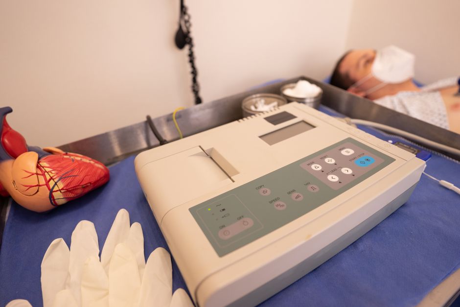
Photo by Los Muertos Crew on Pexels
The rhythmic beating of your heart is called the cardiac cycle, which consists of two phases:
* Systole: The contraction phase, when the ventricles pump blood out of the heart. (e.g., ventricular systole)
* Diastole: The relaxation phase, when the chambers fill with blood. (e.g., ventricular diastole)
These contractions are initiated by the heart's own electrical system, starting with the sinoatrial (SA) node, often called the heart's natural pacemaker.
3. Worked Example
Let's trace a single red blood cell from your big toe back to your big toe.
- Your red blood cell (RBC), now deoxygenated, leaves your toe in a small venule, which merges into larger veins.
- It eventually enters the inferior vena cava.
- From there, it empties into the right atrium.
- It passes through the tricuspid valve into the right ventricle.
- The right ventricle contracts, pushing the RBC through the pulmonary valve into the pulmonary artery.
- The RBC travels to the lungs. Here, it offloads CO2 and picks up O2.
- Now oxygenated, the RBC travels via a pulmonary vein to the left atrium.
- It then passes through the mitral valve into the left ventricle.
- The left ventricle contracts powerfully, propelling the RBC through the aortic valve into the aorta.
- The aorta branches, and the RBC travels down through various arteries to reach your leg and finally returns to your big toe, delivering its oxygen.
4. Key Takeaways
- Your heart is a four-chambered muscular pump vital for circulating blood.
- The right side of the heart handles deoxygenated blood, sending it to the lungs.
- The left side of the heart handles oxygenated blood, pumping it to the rest of the body.
- Valves ensure blood flows in only one direction, preventing backflow.
- The pulmonary circuit oxygenates blood, while the systemic circuit delivers it.
- The cardiac cycle involves systole (contraction) and diastole (relaxation).
Common Mistakes to Avoid

Photo by KATRIN BOLOVTSOVA on Pexels
- Confusing the functions of the right and left sides of the heart; they handle different types of blood.
- Forgetting the role of valves; they're crucial for unidirectional blood flow.
- Mixing up arteries and veins; arteries generally carry blood away from the heart, and veins carry blood towards it (with pulmonary exceptions).
- Thinking blood in the pulmonary artery is oxygenated; it's deoxygenated as it's going to the lungs.
5. Now Try It
Draw a simplified diagram of the heart with its four chambers and the four main valves. Label each chamber and valve, and then use different colored arrows to trace the path of deoxygenated blood (e.g., blue) and oxygenated blood (e.g., red) through the entire heart, indicating where blood leaves for the lungs and where it leaves for the body. You should be able to clearly see the two separate circuits.
Frequently asked about The Human Heart
Study this next
Get the full FSC Biology curriculum
Clone the complete plan to your dashboard for unlimited AI-generated notes, practice quizzes, and a personalised revision schedule.
Save this course free