Detailed Anatomy of the Axial Skeleton
From the HSB curriculum
Detailed Anatomy of the Axial Skeleton
TL;DR
The axial skeleton forms your body's central core, protecting vital organs and providing support. It's made up of the skull, vertebral column, and thoracic cage, each with specific bones and functions. Understanding these components helps you grasp how your body moves and is protected.
1. The Mental Model
Think of the axial skeleton as your body's main structural support beam and protective shell. It’s like the mast of a ship, keeping everything upright, and the walls of a castle, shielding your most important parts. Without it, you'd be a floppy mess and very vulnerable.
2. The Core Material
Your axial skeleton is the central part of your skeletal system, consisting of 80 bones divided into three main regions: the skull, the vertebral column, and the thoracic cage. These bones primarily provide protection for internal organs and a surface for muscle attachment.
The Skull
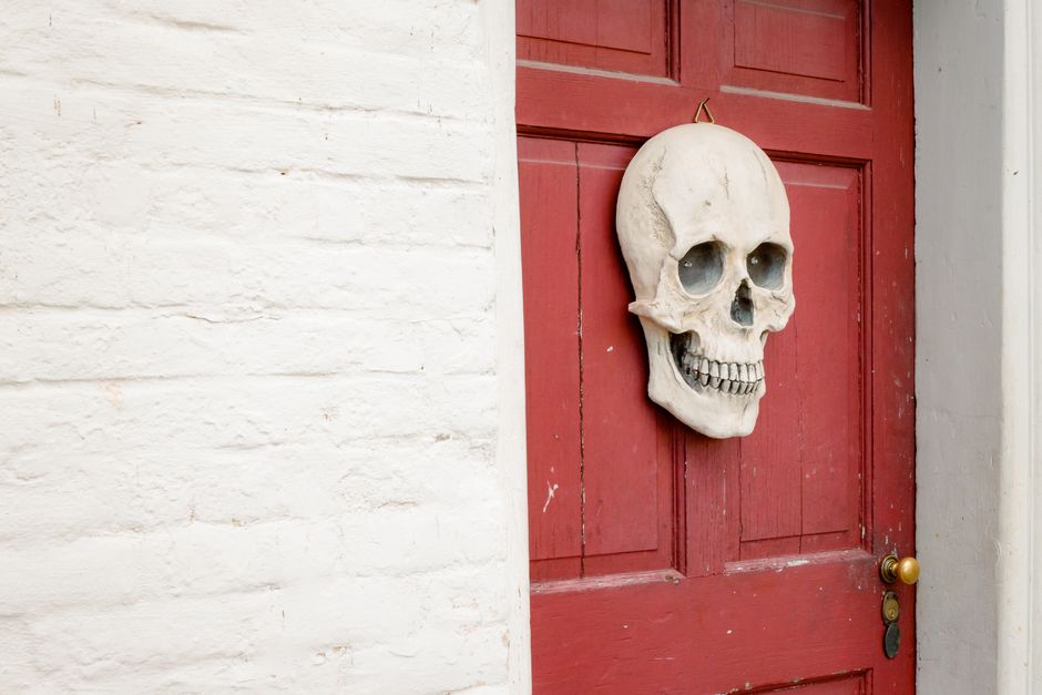
Photo by Michael Morse on Pexels
The skull is a bony structure that forms the head, protecting your brain and housing sensory organs. It's divided into two main parts: the cranium and the facial bones.
- Cranium (8 bones): These bones form the protective case around your brain. They include the frontal, parietal (2), temporal (2), occipital, sphenoid, and ethmoid bones.
- Facial Bones (14 bones): These bones form the structure of your face, support your teeth, and house many sensory organs. Examples are the maxilla (2), mandible, zygomatic (2), and nasal (2) bones. Don't forget the hyoid bone, which is unique as it doesn't articulate with any other bone; it's suspended by ligaments and muscles in your neck.
The Vertebral Column (Spine)

Photo by cottonbro studio on Pexels
Your vertebral column (or spine) extends from the skull to the pelvis, providing support, allowing for movement, and protecting your spinal cord. It's made up of 26 irregular bones called vertebrae, separated by intervertebral discs that act as shock absorbers.
It's divided into five regions:
- Cervical Vertebrae (7): C1-C7, forming your neck. C1 (atlas) and C2 (axis) are specialized for head movement.
- Thoracic Vertebrae (12): T1-T12, forming the upper back. These articulate with your ribs.
- Lumbar Vertebrae (5): L1-L5, forming your lower back. These are the largest and strongest vertebrae.
- Sacrum (1): Five fused vertebrae (S1-S5) that connect to your pelvis.
- Coccyx (1): Your "tailbone," typically made of 3-5 fused vertebrae.
graph TD
    A["Axial Skeleton (80 bones)"] --> B["Skull (22 bones + Hyoid)"]
    A --> C["Vertebral Column (26 bones)"]
    A --> D["Thoracic Cage (25 bones)"]

    B --> B1["Cranium (8 bones)"]
    B --> B2["Facial Bones (14 bones)"]
    B --> B3["Hyoid Bone (1 bone)"]

    C --> C1["Cervical Vertebrae (7)"]
    C --> C2["Thoracic Vertebrae (12)"]
    C --> C3["Lumbar Vertebrae (5)"]
    C --> C4["Sacrum (1)"]
    C --> C5["Coccyx (1)"]

    D --> D1["Sternum (1)"]
    D --> D2["Ribs (12 pairs / 24 total)"]

    C1 --> C1a["Atlas (C1)"]
    C1 --> C1b["Axis (C2)"]

    D2 --> D2a["True Ribs (Pairs 1-7)"]
    D2 --> D2b["False Ribs (Pairs 8-12)"]
    D2b --> D2c["Floating Ribs (Pairs 11-12)"]
The Thoracic Cage
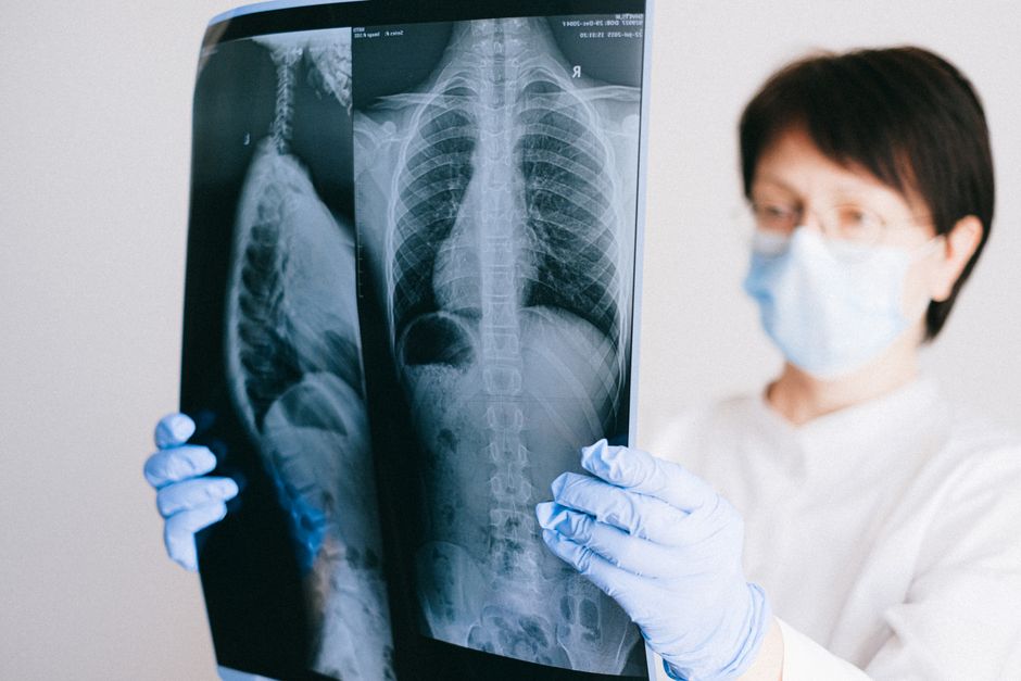
Photo by Anna Shvets on Pexels
The thoracic cage (or rib cage) encloses your chest, protecting your heart and lungs. It's composed of the sternum and ribs.
- Sternum (Breastbone - 1 bone): A flat bone in the center of your chest. It has three parts: the manubrium, body, and xiphoid process.
- Ribs (12 pairs / 24 total bones): These long, curved bones attach to your thoracic vertebrae posteriorly.
- True Ribs (Pairs 1-7): Attach directly to the sternum via costal cartilage.
- False Ribs (Pairs 8-12): Don't directly attach to the sternum. Pairs 8-10 attach indirectly via the cartilage of the rib above them.
- Floating Ribs (Pairs 11-12): Are a type of false rib that doesn't attach to the sternum at all.
3. Worked Example
Let's imagine you're identifying bones on a skeleton model. You find a large, irregular bone with a prominent spine and two large transverse processes that articulates with a rib.
- Is it part of the skull? No, the skull bones are mostly flat or intricately shaped for the head, and wouldn't articulate with a rib in this way.
- Is it part of the thoracic cage? While ribs are in the thoracic cage, this bone is articulating with a rib, meaning it's likely a vertebra. The sternum is flat and central.
- Is it a vertebra? Yes, the description fits a vertebra.
- Which type of vertebra? The articulation with a rib immediately tells you it's a thoracic vertebra. Thoracic vertebrae (T1-T12) are unique among the vertebral regions for having facets (small surfaces) on their bodies and transverse processes where ribs attach. The prominent spine and large transverse processes also align with their role in supporting the rib cage.
Therefore, based on its characteristics and articulations, you've identified it as a thoracic vertebra.
4. Key Takeaways
- The axial skeleton consists of the skull, vertebral column, and thoracic cage.
- The skull protects the brain and houses facial structures and sensory organs.
- The vertebral column supports the body, allows movement, and protects the spinal cord.
- The thoracic cage protects the heart and lungs, facilitating respiration.
- The hyoid bone is unique as it doesn't articulate with any other bone.
-
Ribs are classified as true, false, or floating based on their sternal attachment.
-
Common Mistakes to Avoid:
- Confusing axial skeleton bones with appendicular skeleton bones (limbs, girdles).
- Forgetting the hyoid bone is part of the skull complex.
- Mixing up the number of vertebrae in each spinal region (e.g., thinking there are 10 cervical vertebrae).
- Not understanding why specific bones have their unique shapes (e.g., flat cranial bones for protection, large lumbar vertebrae for weight bearing).
5. Now Try It
Take a pen and paper and, without looking at your notes, sketch and label the main components of the axial skeleton. For each component (skull, vertebral column, thoracic cage), list at least two specific bones and their primary function.
Success looks like: You've correctly drawn the rough outline of each major part (skull, spine, rib cage) and accurately listed two bones for each (e.g., frontal bone, sternum, atlas) along with a correct function (e.g., brain protection, spinal cord protection, heart/lung protection).
Frequently asked about Detailed Anatomy of the Axial Skeleton
More from HSB
Get the full HSB curriculum
Clone the complete plan to your dashboard for unlimited AI-generated notes, practice quizzes, and a personalised revision schedule.
Create Free Account