Introduction to Biological Membranes
From the Anatomy Skin system curriculum
Introduction to Biological Membranes
TL;DR
Biological membranes are flexible barriers that separate the inside of a cell from its outside, or divide a cell into compartments. They're mainly made of lipids and proteins, forming a selectively permeable barrier. This structure allows membranes to control what goes in and out, which is crucial for cell function and survival.
1. The Mental Model
Imagine a cell as a house. The biological membrane is like the walls, roof, and doors – it defines the boundaries, provides structure, and controls who or what comes in and out. It's not a rigid brick wall, though; it's more like a flexible, smart screen.
2. The Core Material
You'll find biological membranes everywhere in a cell. The plasma membrane forms the cell's outer boundary, but there are also membranes inside the cell that create organelles like the nucleus, mitochondria, and endoplasmic reticulum. These internal membranes allow different cellular processes to happen in specific, controlled environments.
2.1. The Lipid Bilayer: The Foundation
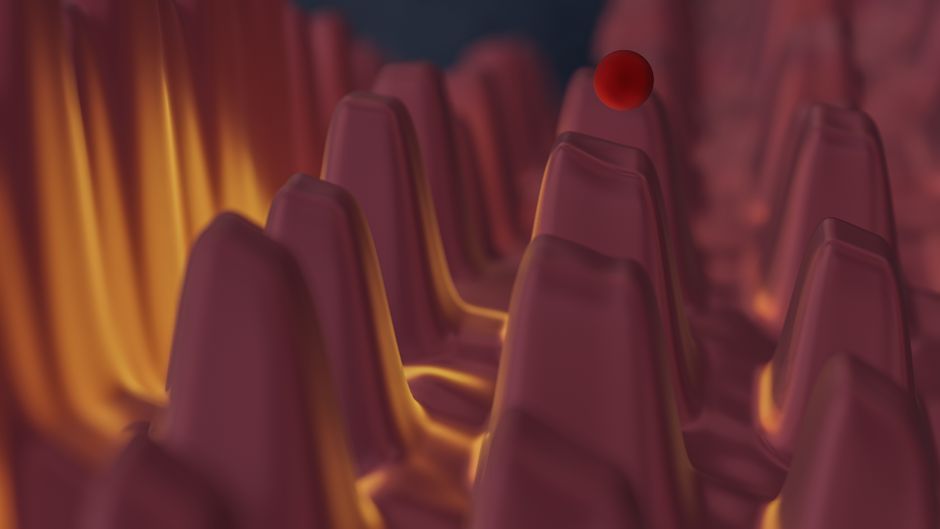
Photo by Steve A Johnson on Pexels
The fundamental structure of any biological membrane is the lipid bilayer. It's essentially two layers of lipid molecules, primarily phospholipids, arranged tail-to-tail.
Think of a phospholipid as having two main parts:
* Hydrophilic head: This part loves water ("hydro" = water, "philic" = loving). It contains a phosphate group and faces outwards, towards the watery environments inside and outside the cell.
* Hydrophobic tails: These are long fatty acid chains that hate water ("hydro" = water, "phobic" = fearing). They face inwards, away from the water, forming the core of the membrane.
This arrangement naturally forms a stable barrier in a watery environment. The hydrophobic tails are protected from water, while the hydrophilic heads interact with it.
graph TD
    H1("Hydrophilic Head") --> T1("Hydrophobic Tails")
    T1 --> T2("Hydrophobic Tails")
    T2 --> H2("Hydrophilic Head")

    subgraph "Phospholipid 1"
        H1
        T1
    end

    subgraph "Phospholipid 2"
        H2
        T2
    end

    L1[("Outside Cell (Watery)")] --- H1
    L2[("Inside Cell (Watery)")] --- H2
     bilayer("Lipid Bilayer Core (Hydrophobic)") --- T1
    bilayer --- T2
2.2. Proteins: The Workers and Communicators

Photo by the northern lense on Pexels
While the lipid bilayer provides the basic structure, proteins are embedded within or attached to it, giving the membrane most of its specific functions.
- Integral proteins: These proteins are embedded directly within the lipid bilayer. Some span the entire membrane (transmembrane proteins), acting as channels or carriers to move substances across. Others are only partially embedded.
- Peripheral proteins: These proteins are loosely attached to the surface of the membrane, either on the inside or outside. They often play roles in cell signaling or enzyme activity.
Membrane proteins do a lot:
* Transport: Moving specific molecules (like glucose or ions) across the membrane.
* Enzymatic activity: Catalyzing reactions at the membrane surface.
* Signal transduction: Receiving chemical messages from outside the cell and relaying them inside.
* Cell-cell recognition: Identifying other cells.
* Intercellular joining: Linking adjacent cells together.
* Attachment to the cytoskeleton and extracellular matrix: Providing structural support and maintaining cell shape.
2.3. Carbohydrates: The Identifiers
Photo by Vinícius Caricatte on Pexels
Short chains of carbohydrates (sugars) are often found attached to lipids (forming glycolipids) or proteins (forming glycoproteins) on the outer surface of the plasma membrane. These form what's called the glycocalyx. They're like cellular ID tags, crucial for cell recognition and adhesion.
3. Worked Example
Let's consider how a cell takes in glucose. Glucose is a polar molecule, meaning it has a slight electrical charge, and it's too large to simply diffuse through the hydrophobic lipid bilayer on its own.
- Recognition: Glucose molecules approach the outer surface of the plasma membrane.
- Binding: A specific integral protein in the membrane, called a glucose transporter, has a binding site for glucose. Glucose binds to this site.
- Conformational Change: The binding of glucose causes the transporter protein to change its shape.
- Transport: This shape change moves the glucose molecule from the outside to the inside of the cell.
- Release: The glucose is released into the cytoplasm, and the transporter protein returns to its original shape, ready to transport another glucose molecule.
This entire process relies on the specific structure and function of the membrane proteins embedded within the lipid bilayer, highlighting the selective permeability and dynamic nature of biological membranes.
4. Key Takeaways
- Biological membranes define cell boundaries and compartmentalize internal cellular activities.
- The core structure is a lipid bilayer formed by phospholipids, with hydrophilic heads facing water and hydrophobic tails forming the inner core.
- Proteins embedded within or attached to the membrane perform most of its specific functions, including transport, signaling, and enzymatic activity.
- Carbohydrates on the outer surface act as cellular identification tags.
- Membranes are selectively permeable, meaning they control what substances can pass through, maintaining a stable internal environment.
Common Mistakes to Avoid:
* Don't think of membranes as rigid, static structures; they're fluid and dynamic.
* Don't forget the role of proteins; the lipid bilayer alone is just a barrier, not a functional membrane.
* Don't confuse the hydrophilic heads with the hydrophobic tails – getting their orientation wrong is a big one.
* Don't assume all molecules can pass through freely; membranes are highly selective.
5. Now Try It
Draw a simple diagram of a small section of a biological membrane. Label the hydrophilic heads, hydrophobic tails, and at least one integral protein and one peripheral protein. Add a carbohydrate chain to either a lipid or a protein on the outer surface. Think about why each part is positioned where it is. What success looks like: Your drawing clearly shows the bilayer structure, the correct orientation of phospholipids, and the placement of proteins and carbohydrates as discussed, indicating an understanding of their basic roles.
Frequently asked about Introduction to Biological Membranes
Get the full Anatomy Skin system curriculum
Clone the complete plan to your dashboard for unlimited AI-generated notes, practice quizzes, and a personalised revision schedule.
Create Free Account