Nervous Tissue: Structure and Signaling
From the Regional terms and Histology curriculum
Nervous Tissue: Structure and Signaling
TL;DR
Nervous tissue, made of neurons and glial cells, is how your body sends fast electrical and chemical messages. Neurons generate electrical signals called action potentials to communicate, while glial cells support them. This intricate system allows for sensation, thought, and movement.
1. The Mental Model
Think of your nervous system as a vast, complex electrical grid. Neurons are the long wires carrying signals, and glial cells are the maintenance crew keeping everything running smoothly. Signals travel rapidly, letting different parts of your body talk to each other instantly.
2. The Core Material
Your nervous system is the master control and communication system of your body. It allows you to sense the world, think, move, and regulate internal functions. It's made of two main types of cells: neurons and neuroglia (or glial cells).
2.1. Neurons: The Communicators
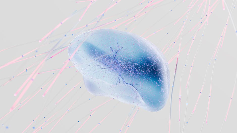
Photo by Google DeepMind on Pexels
Neurons are the fundamental units of the nervous system, specialized for transmitting electrical signals. They have a unique structure:
- Cell Body (Soma): This is the neuron's main part, containing the nucleus and most organelles. It's where the neuron's "decisions" are made.
- Dendrites: These are short, branching extensions that receive incoming signals from other neurons. Think of them as the neuron's "antennas."
- Axon: A single, long extension that carries electrical signals (action potentials) away from the cell body to other neurons, muscles, or glands. Axons can be very long, sometimes over a meter!
- Axon Terminals: The very end of the axon, where it branches out. These terminals release chemical messengers called neurotransmitters into the synaptic cleft.
- Myelin Sheath: A fatty insulating layer that wraps around many axons. It's formed by glial cells and dramatically speeds up signal transmission. Gaps in the myelin are called Nodes of Ranvier.
2.2. Neuroglia (Glial Cells): The Support Crew
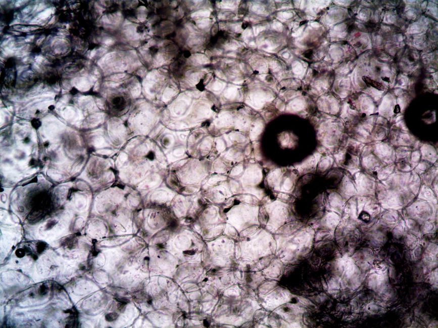
Photo by turek on Pexels
Glial cells don't transmit signals themselves, but they are crucial for neuron function and survival. There are several types:
- Astrocytes: Found in the Central Nervous System (CNS - brain and spinal cord). They support neurons, regulate the chemical environment, and form the blood-brain barrier.
- Oligodendrocytes: Also in the CNS. They produce myelin sheaths for axons in the brain and spinal cord.
- Microglia: Small, mobile immune cells in the CNS. They act as the nervous system's clean-up crew, removing debris and pathogens.
- Ependymal Cells: Line the cavities of the brain and spinal cord, producing and circulating cerebrospinal fluid (CSF).
- Schwann Cells: Found in the Peripheral Nervous System (PNS - nerves outside the brain and spinal cord). They form myelin sheaths for axons in the PNS.
- Satellite Cells: Surround neuron cell bodies in the PNS, providing support and nutrient regulation.
2.3. How Neurons Signal: The Action Potential
Photo by Google DeepMind on Pexels
Neuronal signaling is primarily electrical, involving rapid changes in the electrical charge across the neuron's membrane. This electrical signal is called an action potential.
- Resting Membrane Potential: A neuron at rest has a negative charge inside compared to outside (around -70mV). This is maintained by ion pumps (like the Na+/K+ pump) and ion channels.
- Threshold Stimulus: If dendrites receive enough excitatory signals, the membrane potential at the axon hillock (where the axon leaves the cell body) becomes less negative, reaching a "threshold" (e.g., -55mV).
- Depolarization: Voltage-gated sodium (Na+) channels open rapidly, allowing Na+ ions to rush into the cell. The inside of the cell becomes positive. This is the rising phase of the action potential.
- Repolarization: Voltage-gated sodium channels inactivate, and voltage-gated potassium (K+) channels open more slowly, allowing K+ ions to flow out of the cell. The inside of the cell becomes negative again.
- Hyperpolarization (Undershoot): K+ channels close slowly, leading to a brief period where the membrane potential is even more negative than the resting potential.
- Restoration: The Na+/K+ pump works to restore the original ion distribution, returning the neuron to its resting membrane potential.
Action potentials are "all-or-nothing" events; they either happen completely or not at all. Once triggered, they propagate down the axon without losing strength. Myelin speeds up this propagation by allowing the signal to "jump" between Nodes of Ranvier (saltatory conduction).
graph TD
    A["Resting State (-70mV)"] --> B{"Stimulus Reaches Threshold (-55mV)?"}
    B -- Yes --> C["Depolarization (Na+ influx)"]
    C --> D["Repolarization (K+ efflux)"]
    D --> E["Hyperpolarization (K+ channels close slowly)"]
    E --> F["Return to Resting State"]
    B -- No --> A
2.4. Synaptic Transmission: Chemical Communication

Photo by Ron Lach on Pexels
When an action potential reaches the axon terminal, it triggers the release of neurotransmitters into the synaptic cleft (the tiny gap between neurons). These neurotransmitters bind to receptors on the dendrite or cell body of the next neuron (the postsynaptic neuron), causing either excitation (making it more likely to fire an action potential) or inhibition (making it less likely). This chemical step is how signals jump from one neuron to the next.
3. Worked Example
Let's trace a simple signal: touching a hot stove.
- Stimulus: Your finger touches the hot stove. Heat receptors in your skin are activated.
- Sensory Neuron Activation: These receptors generate a signal that reaches the dendrites of a sensory neuron in your finger. If the stimulus is strong enough, it triggers an action potential in that sensory neuron.
- Signal Propagation: The action potential travels rapidly down the sensory neuron's axon (which is likely myelinated, speeding it up) towards your spinal cord.
- Synaptic Transmission (Spinal Cord): At the spinal cord, the sensory neuron's axon terminals release neurotransmitters (e.g., glutamate) into a synapse with an interneuron.
- Interneuron Activation: These neurotransmitters bind to receptors on the interneuron, generating an action potential in the interneuron.
- Synaptic Transmission (Motor Neuron): The interneuron then synapses with a motor neuron. It releases neurotransmitters that excite the motor neuron.
- Motor Neuron Activation: The motor neuron generates an action potential, which travels down its axon to the muscles in your arm.
- Muscle Contraction: At the neuromuscular junction, the motor neuron releases acetylcholine, causing your arm muscles to contract and pull your hand away. All of this happens almost instantaneously!
4. Key Takeaways
- Neurons are specialized for rapid electrical signal transmission via action potentials.
- Glial cells provide essential support, insulation, and maintenance for neurons.
- The action potential is an "all-or-nothing" electrical event caused by ion movement across the neuron membrane.
- Myelin sheaths, formed by glial cells, significantly increase the speed of action potential conduction.
- Synapses are where neurons chemically communicate by releasing neurotransmitters.
- The nervous system is broadly divided into the Central Nervous System (CNS) and Peripheral Nervous System (PNS).
- Different glial cell types have specific roles and locations (e.g., Schwann cells in PNS, oligodendrocytes in CNS).
Common mistakes to avoid:
- Confusing the roles of neurons (signaling) and glial cells (support).
- Thinking action potentials are graded; they are always the same strength once triggered.
- Forgetting that the resting membrane potential is negative inside the neuron.
- Mixing up which glial cells produce myelin in the CNS versus the PNS.
5. Now Try It
Imagine you're explaining how a reflex arc works (like the hot stove example) to a friend. On a piece of paper, draw a simplified diagram showing at least three neurons (sensory, interneuron, motor) and label their key parts (dendrites, cell body, axon, axon terminal). For each synapse, briefly describe what's happening in terms of neurotransmitter release and receptor binding.
Success looks like: A clear, labeled diagram with arrows showing signal direction, and short explanations at each synapse that correctly identify neurotransmitters being released and acting on the next neuron.
Frequently asked about Nervous Tissue: Structure and Signaling
More from Regional terms and Histology
Get the full Regional terms and Histology curriculum
Clone the complete plan to your dashboard for unlimited AI-generated notes, practice quizzes, and a personalised revision schedule.
Create Free Account