Surgical Anatomy, Physiology, and Pathology
From the Surgery curriculum
TL;DR
Understanding surgical anatomy, physiology, and pathology is crucial for safe and effective surgery. You'll learn the normal structures and functions, how diseases alter them, and how surgical interventions impact these systems. This integrated knowledge helps you anticipate challenges, perform precise procedures, and manage patient outcomes.
1. The Mental Model
Think of it like being a mechanic: you need to know how a car's engine should work (physiology), what each part is (anatomy), and what happens when parts break or get clogged (pathology) to fix it properly. In surgery, the patient is the "machine," and you're the "mechanic."
2. The Core Material
When you're approaching surgery, you're not just cutting and sewing; you're interacting with a living system. This means you need a deep understanding of three interconnected areas:
Anatomical Foundations

Photo by Magda Ehlers on Pexels
This is about "where things are." You need to know the precise location, relationships, and structural details of organs, vessels, nerves, and tissues. For instance, knowing the exact course of the recurrent laryngeal nerve during thyroid surgery is critical to prevent vocal cord paralysis. It's not just memorization; it's visualizing structures in 3D and understanding their variations.
Physiological Principles
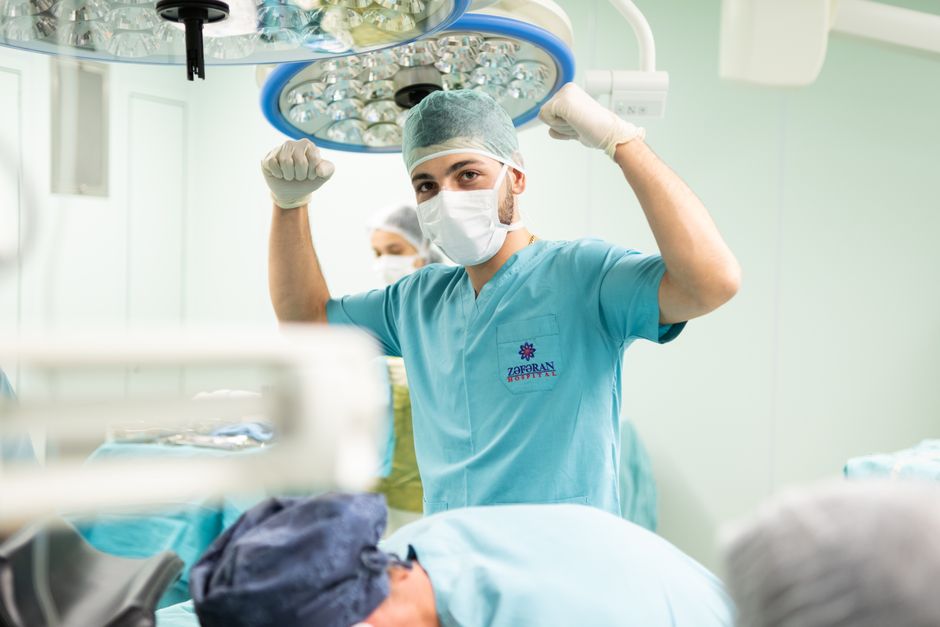
Photo by Javid Hashimov on Pexels
This is about "how things work." You need to understand the normal functions of organs and systems. How does the liver metabolize drugs? How does the heart pump blood? How does the kidney regulate fluids? This knowledge guides your pre-operative assessment (e.g., assessing cardiac risk), intra-operative decisions (e.g., managing blood loss), and post-operative care (e.g., monitoring kidney function).
Pathological Processes

Photo by Muhammad Khawar Nazir on Pexels
This is about "what goes wrong." You need to understand how disease alters normal anatomy and physiology. A tumor changes the structure of an organ and impairs its function. Inflammation can make tissues fragile and bleed easily. Ischemia can lead to tissue death. Knowing the pathology helps you understand why symptoms occur, predict the extent of disease, and choose the most appropriate surgical approach.
The Interplay
Photo by Javid Hashimov on Pexels
These three areas aren't separate; they're constantly influencing each other. Here's a simple flow:
graph LR
    A["Normal Anatomy (Structure)"] --> B["Normal Physiology (Function)"]
    B --> C["Disease Process (Pathology)"];
    C --> D["Altered Anatomy"];
    C --> E["Altered Physiology"];
    D --> F["Surgical Intervention"];
    E --> F;
    F --> G["Post-Op Anatomy/Physiology"];
    G --> H["Patient Outcome"];
For example, in appendicitis:
* Anatomy: The appendix is a small, finger-shaped pouch projecting from your large intestine.
* Physiology: Its exact function isn't fully clear, but it's part of the gut.
* Pathology: Obstruction (e.g., by a fecalith) leads to inflammation (appendicitis), increasing pressure, reducing blood flow, and potentially causing rupture.
* Surgical Intervention: Appendectomy involves carefully dissecting the inflamed appendix, ligating its blood supply, and removing it, ensuring no spillage of pus.
* Post-Op: The patient recovers, and the body adapts to the absence of the appendix.
3. Worked Example
Let's consider a patient with a gallstone obstructing the common bile duct (choledocholithiasis).
- Anatomy: You recall the biliary tree: liver produces bile, bile ducts merge into common hepatic duct, which joins the cystic duct (from the gallbladder) to form the common bile duct (CBD). The CBD typically empties into the duodenum at the ampulla of Vater, often co-joining with the pancreatic duct. You visualize the pancreas, duodenum, and major vessels like the portal vein and hepatic artery nearby.
- Physiology: Bile aids in fat digestion. When the CBD is obstructed, bile backs up. Pancreatic enzymes can also back up if the stone is at the ampulla.
- Pathology:
- Obstruction: The gallstone blocks bile flow.
- Jaundice: Bilirubin, a component of bile, builds up in the blood, leading to yellow skin/eyes.
- Cholangitis: Stagnant bile can get infected, leading to severe infection, fever, and sepsis.
- Pancreatitis: If the stone impacts the pancreatic duct opening, pancreatic enzymes can auto-digest the pancreas.
- Anatomical Changes: The bile ducts above the obstruction will dilate (get wider). The gallbladder might also be inflamed (cholecystitis).
- Surgical Implications: Your goals are to relieve the obstruction and manage any infection. You might perform an Endoscopic Retrograde Cholangiopancreatography (ERCP) to retrieve the stone, or a surgical exploration of the CBD (CBD exploration) to remove the stone, often followed by a cholecystectomy (gallbladder removal) to prevent recurrence. During surgery, you'd be very mindful of the anatomy of the structures in Calot's triangle (cystic duct, common hepatic duct, cystic artery) to avoid injuring critical structures.
4. Key Takeaways
- Surgical anatomy defines the "roadmap" for your procedures, guiding your incisions and dissections.
- Physiology explains "how the body works normally" and "what goes wrong" when disease strikes.
- Pathology details the specific changes caused by disease, impacting surgical approach and prognosis.
- These three areas are interconnected; a change in one often affects the others.
- Understanding variations in anatomy is crucial to avoid iatrogenic injury.
- Predicting physiological responses to surgical stress helps in perioperative management.
-
Knowing the natural history of a disease helps you tailor the extent of your surgical intervention.
-
Common mistakes to avoid:
- Relying solely on textbooks; always consider individual patient variations.
- Underestimating the physiological impact of surgical stress on compromised patients.
- Not correlating imaging findings with your anatomical and pathological knowledge.
- Neglecting to re-evaluate the pathology during surgery (e.g., unexpectedly finding a tumor instead of inflammation).
5. Now Try It
Take a common surgical condition you're familiar with, like an inguinal hernia. For about 15 minutes, outline the following:
1. Anatomy: What layers do you cut through? What are the key structures in the inguinal canal? What's Hesselbach's triangle?
2. Physiology: How does increased intra-abdominal pressure relate to hernia formation? What are the physiological consequences of bowel strangulation?
3. Pathology: How does a direct hernia differ from an indirect hernia? What are the pathological changes in an incarcerated or strangulated hernia?
4. Surgical Implications: What anatomical landmarks guide your repair? What physiological changes do you monitor during and after repair?
Success looks like clearly articulating these points without looking them up, demonstrating your integrated understanding of how these three core components apply to a real-world surgical problem.
Frequently asked about Surgical Anatomy, Physiology, and Pathology
Study this next
Get the full Surgery curriculum
Clone the complete plan to your dashboard for unlimited AI-generated notes, practice quizzes, and a personalised revision schedule.
Save this course free