Introduction to Ultrasound Transducers
From the sonography curriculum
Introduction to Ultrasound Transducers
TL;DR
Ultrasound transducers are the heart of sonography, converting electrical energy into sound waves and back again to create images. They use piezoelectric crystals that vibrate when an electric current is applied and generate a current when vibrated by sound. Different transducer types are chosen based on the body part being scanned, as they have unique frequencies and beam shapes.
1. The Mental Model
Think of a transducer as a microphone and a speaker combined, but for very high-pitched sound. It sends out sound waves and then listens for the echoes, painting a picture of what's inside.
2. The Core Material
Ultrasound transducers are essentially sophisticated antennas for sound waves. They're critical because they perform two main functions:
1. Transmission: They convert electrical energy into high-frequency sound waves (ultrasound) that penetrate the body.
2. Reception: They capture the reflected sound waves (echoes) and convert them back into electrical signals for the ultrasound machine to process into an image.
This conversion magic happens thanks to piezoelectric crystals within the transducer. When you apply an electric current to these crystals, they rapidly change shape and vibrate, generating sound waves. Conversely, when sound waves hit these crystals, they deform and produce a tiny electric current.
Transducers come in various shapes and sizes, each designed for specific clinical applications. Their main differences lie in:
- Frequency: This determines how deep the sound waves can go and how detailed the image will be.
- High frequency (e.g., 7-18 MHz): Provides excellent resolution (clearer, more detailed images) but has poor penetration (can't go very deep). Ideal for superficial structures like thyroid, breast, and vessels.
- Low frequency (e.g., 1-5 MHz): Offers good penetration (can go deep) but has poor resolution (less detailed images). Used for deeper organs like the liver, kidneys, and during abdominal scans.
- Footprint/Shape: The part of the transducer that touches the patient. This influences the shape of the sound beam and the field of view.
Types of Transducers
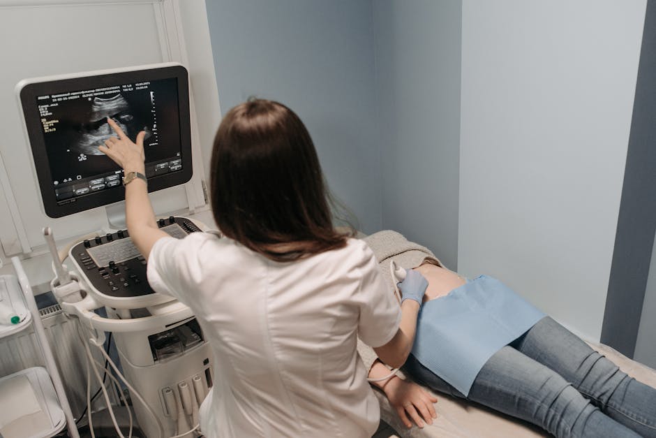
Photo by Pavel Danilyuk on Pexels
There are three primary types you'll encounter:
Linear Array Transducer
- Beam Shape: Rectangular (straight lines of sound).
- Frequency Range: Typically high (7-18 MHz).
- Use: Superficial structures (e.g., thyroid, breast, vascular access, musculoskeletal). Gives a wide, flat view.
Curvilinear (Convex) Array Transducer
- Beam Shape: Curved, fan-shaped (like looking through a fisheye lens).
- Frequency Range: Typically low (1-5 MHz).
- Use: Deeper structures (e.g., abdominal, obstetric, gynecological). Allows a wider field of view for larger areas.
Phased Array (Sector) Transducer
- Beam Shape: Narrow at the top, widening into a sector/pie shape.
- Frequency Range: Low to medium (2-7 MHz).
- Use: Structures requiring penetration through a small acoustic window (e.g., cardiac, transcranial). It's great for getting between ribs.
graph TD
    A["Ultrasound Transducer"] --> B["Piezoelectric Crystals"];
    B --> C["Electrical Energy In"];
    C --> D["Crystal Vibration (Sound Out)"];
    B --> E["Sound Waves Reflecting Back"];
    E --> F["Crystal Vibration (Electrical Signal Out)"];
    F --> G["Image Processing Unit"];

    subgraph Transducer Types
        H["Linear Array"] --> H1["High Frequency"];
        H1 --> H2["Rectangular Beam"];
        H2 --> H3["Superficial Organs (e.g., Thyroid, Breast)"];

        I["Curvilinear Array"] --> I1["Low Frequency"];
        I1 --> I2["Curved, Fan-shaped Beam"];
        I2 --> I3["Deep Organs (e.g., Abdomen, OB/GYN)"];

        J["Phased Array"] --> J1["Low/Medium Frequency"];
        J1 --> J2["Sector/Pie-shaped Beam"];
        J2 --> J3["Cardiac, Transcranial (Small Acoustic Window)"];
    end

    A --- H;
    A --- I;
    A --- J;
3. Worked Example
Imagine you're asked to perform an ultrasound on a patient's carotid artery to check for plaque.
- Clinical Need: Visualize a superficial blood vessel with high detail to identify small abnormalities.
- Transducer Selection Criteria: You need high resolution because the artery is close to the skin and you're looking for fine details like plaque. Penetration isn't a major concern.
- Choice: You'd select a linear array transducer.
- Why? It operates at a high frequency (e.g., 10-15 MHz), providing excellent resolution for superficial structures. Its rectangular footprint gives you a broad, uncurved view of the artery.
- Application: You'd apply gel to the patient's neck over the carotid artery, place the linear transducer gently, and begin scanning, adjusting the depth and focus to get the clearest image of the vessel walls and lumen.
Now, if the same patient needed an ultrasound of their liver (a deep abdominal organ), you'd switch to a curvilinear array transducer. Why? Because its low frequency (e.g., 3.5 MHz) allows sound waves to penetrate much deeper into the abdomen, even though the image resolution won't be as sharp as for the carotid artery.
4. Key Takeaways
- Transducers convert electrical energy to sound and back using piezoelectric crystals.
- Frequency dictates both image resolution (detail) and penetration depth.
- High frequency means high resolution, shallow penetration; low frequency means low resolution, deep penetration.
- Linear transducers are best for superficial structures due to their high frequency and rectangular field of view.
- Curvilinear transducers are best for deep structures due to their low frequency and wide, fan-shaped field of view.
- Phased array transducers are used when you need to image deep structures through a small "window," like the ribs for the heart.
Common Mistakes to Avoid:
- Don't use a low-frequency transducer for superficial structures; you'll get a blurry image.
- Don't expect a high-frequency transducer to image deep organs; the sound won't penetrate enough.
- Forgetting to apply ultrasound gel; it's essential for sound wave transmission.
- Pressing too hard with the transducer can compress structures and distort the image.
5. Now Try It
Imagine you need to perform an ultrasound on a pregnant patient in their third trimester to assess fetal growth.
1. What type of transducer would you most likely choose?
2. Explain why that transducer is the best choice, specifically mentioning its frequency characteristics and beam shape in relation to the anatomy you're scanning.
Success looks like correctly identifying the transducer type and clearly articulating how its technical specifications (frequency, beam shape) match the clinical requirement of imaging a deep structure like a fetus.
Frequently asked about Introduction to Ultrasound Transducers
Get the full sonography curriculum
Clone the complete plan to your dashboard for unlimited AI-generated notes, practice quizzes, and a personalised revision schedule.
Create Free Account