Introduction to Cells and Microscopy
From the Chapter 8: Cells and Cell Functions curriculum
Introduction to Cells and Microscopy
TL;DR
Cells are the fundamental building blocks of all living things, too tiny to see with your naked eye. We use microscopes to magnify them and understand their incredibly complex structures. Learning about cells starts with understanding how we visualize them.
1. The Mental Model
Think of cells like tiny LEGO bricks that make up everything alive. We need special magnifying glasses, called microscopes, to see these tiny bricks and how they're arranged.
2. The Core Material
What Are Cells?

Photo by Monstera Production on Pexels
Cells are the smallest units of life that can perform all essential life processes. Every living organism, from a single bacterium to a massive whale, is made of cells. They come in many shapes and sizes, but they all share basic characteristics like having a boundary (cell membrane), genetic material (DNA), and cellular machinery.
There are two main types of cells:
- Prokaryotic Cells: These are simpler cells, typically single-celled organisms like bacteria. They don't have a nucleus or other membrane-bound organelles. Their genetic material floats freely in the cytoplasm.
- Eukaryotic Cells: These are more complex cells found in animals, plants, fungi, and protists. They have a true nucleus that houses their genetic material, and various specialized, membrane-bound compartments called organelles that perform specific jobs.
Why Do We Need Microscopes?

Photo by Mikhail Nilov on Pexels
Cells are usually too small to be seen with the unaided human eye. Most animal cells are between 10 and 100 micrometers (µm) in diameter, and plant cells are similar or a bit larger. A micrometer is one-millionth of a meter! To study these tiny structures, we need tools that can magnify them significantly. That's where microscopes come in.
Types of Microscopes
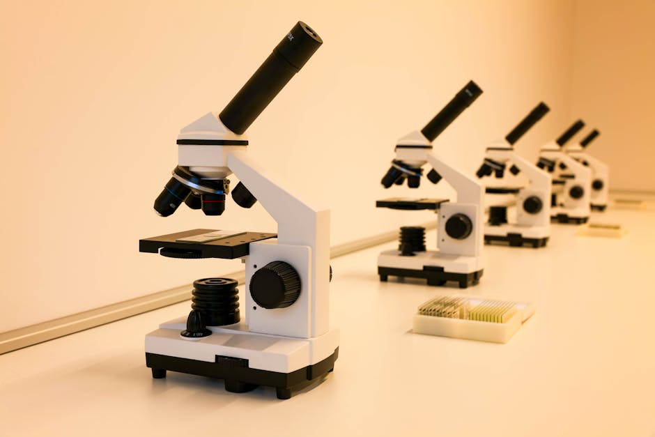
Photo by Vladimir Srajber on Pexels
There are two primary categories of microscopes you'll encounter:
- Light Microscopes (LM): These use visible light and lenses to magnify specimens. They're common in classrooms and labs.
- Compound Light Microscopes: These use multiple lenses for high magnification and are great for viewing living cells and basic cell structures. They can magnify up to about 1000 times.
- Stereo (Dissecting) Microscopes: These provide a 3D view at lower magnifications, useful for observing larger specimens or doing dissections.
- Electron Microscopes (EM): These use beams of electrons instead of light, allowing for much higher magnification and resolution. You can see incredibly fine details with these.
- Scanning Electron Microscopes (SEM): These create detailed 3D images of the surface of a specimen.
- Transmission Electron Microscopes (TEM): These allow you to view internal structures of cells, passing electrons through very thin slices of the specimen.
Here's how we decide which microscope to use:
graph TD
    A["What do you want to see?"] --> B{"Is it alive?"};
    B -- "Yes" --> C{"How big is it/what detail?"};
    B -- "No" --> D{"How big is it/what detail?"};

    C -- "Small (cell structures)" --> LM["Compound Light Microscope"];
    C -- "Larger (small organisms/tissues)" --> SM["Stereo Microscope"];

    D -- "Surface features (high detail)" --> SEM["Scanning Electron Microscope"];
    D -- "Internal structures (ultra-high detail)" --> TEM["Transmission Electron Microscope"];
    D -- "Basic observation (preserved)" --> LM;
Magnification and Resolution
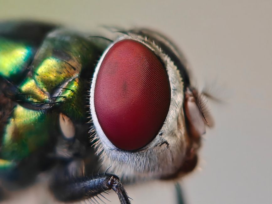
Photo by Sai Pixels on Pexels
- Magnification: This is how much larger an object appears compared to its actual size. If a microscope has a 10x eyepiece and a 40x objective lens, the total magnification is 400x (10 * 40).
- Resolution: This is the ability to distinguish two separate objects as distinct. High resolution means you can see fine details clearly. A blurry, highly magnified image isn't very useful; good resolution is key.
Electron microscopes have much higher resolution than light microscopes because electron beams have a much shorter wavelength than visible light. This allows them to "see" much smaller details.
3. Worked Example
Imagine you've collected a sample of pond water and want to identify any single-celled organisms.
- Preparation: You'd place a small drop of pond water on a microscope slide and cover it with a coverslip.
- Microscope Choice: Since you're looking for living, single-celled organisms and want to observe their general movement and shape, a compound light microscope is your best bet. An electron microscope would kill the organisms and require extensive preparation.
- Observation: You start with a low power objective lens (e.g., 4x or 10x) to get a wide field of view and locate interesting areas.
- Magnification Adjustment: Once you spot something, you switch to a higher power objective lens (e.g., 40x) to see more detail. If your eyepiece is 10x, your total magnification at this point would be 10x * 40x = 400x.
- Focusing: You adjust the fine focus knob to bring the organisms into sharp view, revealing their internal structures (if visible) and how they move. You might identify an Amoeba or Paramecium based on its shape and movement.
4. Key Takeaways
- Cells are the basic structural and functional units of all living things.
- There are two main cell types: prokaryotic (simple, no nucleus) and eukaryotic (complex, with a nucleus and organelles).
- Most cells are microscopic and require special tools to be seen.
- Light microscopes use light to magnify specimens, suitable for living cells and general structures.
- Electron microscopes use electron beams for much higher magnification and resolution, revealing ultra-fine details.
- Magnification is how much larger an image appears, while resolution is the clarity or ability to distinguish separate objects.
- Different microscopes are chosen based on what you want to observe (living vs. dead, surface vs. internal, level of detail).
Common mistakes to avoid:
- Don't confuse magnification with resolution; high magnification without good resolution just gives a bigger blur.
- Don't try to use an electron microscope on living samples; they require specimens to be dead and specially prepared.
- Forgetting to focus properly at each magnification level on a light microscope will lead to blurry images.
- Assuming all cells are the same; they vary greatly in size, shape, and complexity.
5. Now Try It
Spend 15 minutes researching a specific type of cell (e.g., a neuron, a plant leaf cell, a bacterium). Describe its typical size, key features, and explain which type of microscope you would use to observe its internal organelles versus its surface structure, and why. What would be the approximate total magnification you'd aim for in each case?
Frequently asked about Introduction to Cells and Microscopy
Study this next
Get the full Chapter 8: Cells and Cell Functions curriculum
Clone the complete plan to your dashboard for unlimited AI-generated notes, practice quizzes, and a personalised revision schedule.
Create Free Account