Anatomical Foundations of the Head and Face
From the Head, Neck, and Lymph curriculum
Anatomical Foundations of the Head and Face
TL;DR
The head and face are complex structures, built upon a bony skull that protects the brain and forms the facial framework. Key soft tissues, including muscles, nerves, and vessels, enable movement, sensation, and essential functions like chewing and facial expression. Understanding these layers is crucial for clinical applications and recognizing potential issues.
1. The Mental Model
Think of your head and face like a house: the skull is the sturdy frame, muscles are the movable parts like doors and windows, and nerves and blood vessels are the electrical and plumbing systems connecting everything. Each part has a specific job, and they all work together seamlessly.
2. The Core Material
The head and face are incredibly intricate, housing vital organs and performing essential functions. To understand them, we'll break them down into their primary components: the cranium (skull) and the various soft tissues that cover and fill it.
The Cranium: Your Bony Protector
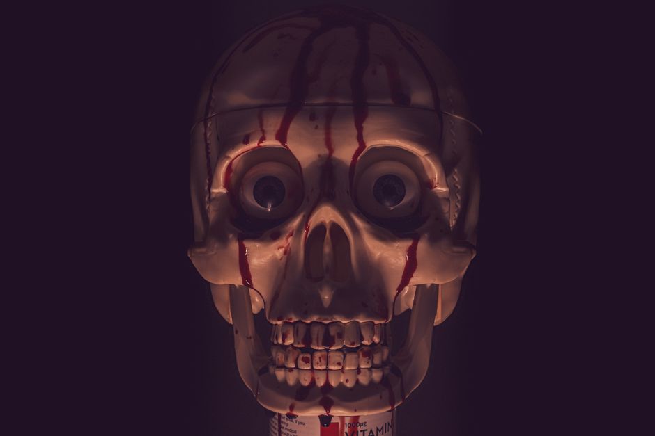
Photo by Boys in Bristol Photography on Pexels
The skull is a complex bony structure that serves several key purposes: it protects the brain, supports the facial structures, and provides attachment points for muscles. It's not one solid bone but a collection of 22 bones (mostly fused in adults) divided into two main parts:
- Neurocranium: This forms the protective casing around the brain. It includes bones like the frontal, parietal, temporal, and occipital bones.
- Viscerocranium (Facial Skeleton): These bones form the face and support organs like the eyes and teeth. Important bones here are the zygomatic (cheekbones), maxilla (upper jaw), mandible (lower jaw), and nasal bones.
Soft Tissues: Movement, Sensation, and Supply

Photo by Funkcinės Terapijos Centras on Pexels
Layered over and within the bony framework are the soft tissues that bring the head and face to life.
Muscles
Muscles are responsible for all movement. In the head and face, they're broadly categorized:
- Muscles of Mastication: These are powerful muscles (temporalis, masseter, medial pterygoid, lateral pterygoid) that move the mandible for chewing.
- Muscles of Facial Expression: These muscles, like the orbicularis oculi (around the eye) and orbicularis oris (around the mouth), are thinner and attach directly to skin, allowing for a wide range of expressions.
- Neck Muscles: While not strictly facial, muscles like the sternocleidomastoid and trapezius are crucial for head movement and posture.
Nerves
Nerves are the communication network, carrying signals for sensation and movement.
- Cranial Nerves: Twelve pairs of nerves emerge directly from the brain. Several are critical for the head and face:
- Trigeminal Nerve (CN V): The primary sensory nerve of the face and motor nerve for muscles of mastication. It has three main branches: ophthalmic, maxillary, and mandibular.
- Facial Nerve (CN VII): Primarily motor, controlling all muscles of facial expression. It also carries taste sensation.
- Vagus Nerve (CN X): Plays a role in speech and swallowing, among many other functions.
- Spinal Nerves: Contribute to sensation and movement in the back of the head and neck.
Blood Supply
An extensive network of arteries and veins ensures the head and face receive oxygenated blood and remove waste.
- Arteries: The common carotid artery on each side branches into the internal carotid (supplying the brain and eyes) and the external carotid (supplying the face, scalp, and neck). Key branches of the external carotid include the facial artery and superficial temporal artery.
- Veins: Veins generally follow a similar path to arteries, draining deoxygenated blood. The internal jugular vein is the main drainage pathway from the head and neck back to the heart.
graph TD
    A["Head & Face (Anatomical Foundations)"] --> B["Bony Framework (Cranium)"]
    A --> C["Soft Tissues"]

    B --> D["Neurocranium (Brain Protection)"]
    B --> E["Viscerocranium (Facial Skeleton)"]

    C --> F["Muscles"]
    C --> G["Nerves"]
    C --> H["Blood Vessels"]

    D --> D1["Frontal Bone"]
    D --> D2["Parietal Bone"]
    D --> D3["Temporal Bone"]
    D --> D4["Occipital Bone"]

    E --> E1["Zygomatic Bone (Cheek)"]
    E --> E2["Maxilla (Upper Jaw)"]
    E --> E3["Mandible (Lower Jaw)"]
    E --> E4["Nasal Bones"]

    F --> F1["Muscles of Mastication"]
    F --> F2["Muscles of Facial Expression"]
    F --> F3["Neck Muscles"]

    G --> G1["Cranial Nerves"]
    G --> G2["Spinal Nerves"]

    H --> H1["Arteries"]
    H --> H2["Veins"]

    G1 --> G1a["Trigeminal Nerve (CN V)"]
    G1 --> G1b["Facial Nerve (CN VII)"]
    G1 --> G1c["Vagus Nerve (CN X)"]

    H1 --> H1a["Common Carotid Artery"]
    H1a --> H1b["Internal Carotid"]
    H1a --> H1c["External Carotid"]
    H1c --> H1d["Facial Artery"]
    H1c --> H1e["Superficial Temporal Artery"]

    H2 --> H2a["Internal Jugular Vein"]
3. Worked Example
Imagine a patient who has experienced trauma to the side of their face, resulting in difficulty chewing and a drooping mouth on one side. Let's trace the potential anatomical structures involved.
The difficulty chewing immediately points to the muscles of mastication. If these muscles (like the masseter or temporalis) are damaged or their nerve supply is compromised, the patient won't be able to bite or grind food effectively. The nerve responsible for innervating these muscles is the Trigeminal Nerve (CN V), specifically its mandibular branch. Trauma in this area could have injured this nerve directly or affected the muscles themselves.
The drooping mouth on one side is a classic sign of damage to the Facial Nerve (CN VII). This nerve controls all the muscles of facial expression, including those around the mouth (like the orbicularis oris and zygomaticus muscles). If this nerve is injured, the muscles it supplies become paralyzed, leading to the characteristic facial asymmetry, difficulty smiling, and sometimes problems with speech and retaining saliva. The external carotid artery or its branches, like the facial artery, would also be at risk in trauma to this area, potentially leading to significant bleeding.
4. Key Takeaways
- The skull's neurocranium protects the brain, while the viscerocranium forms the facial structure and supports sense organs.
- Muscles of mastication (Trigeminal nerve) control chewing, and muscles of facial expression (Facial nerve) enable communication.
- Cranial nerves V (Trigeminal) and VII (Facial) are paramount for facial sensation, chewing, and expression.
- The common carotid artery is the main arterial supply to the head and face, branching into internal and external carotids.
- The internal jugular vein is the primary drainage pathway for deoxygenated blood from the head and neck.
Common Mistakes to Avoid:
- Confusing the roles of the Trigeminal and Facial nerves; one is primarily sensory and mastication, the other for facial expression.
- Forgetting that the skull isn't one solid bone, but several fused bones with specific names.
- Underestimating the complexity and density of structures in relatively small areas of the face.
- Not considering both motor and sensory components when assessing facial injuries or conditions.
5. Now Try It
Spend 15 minutes reviewing a labeled diagram of the human skull, focusing on the names and general locations of the major neurocranium and viscerocranium bones. Then, try to identify the major muscles of mastication and facial expression, and trace the general path of the Trigeminal and Facial nerves. What would happen if the temporal bone was fractured, and which key structures might be immediately at risk? What would success look like? You'd be able to correctly label at least 8-10 major skull bones and describe the basic function of the Trigeminal and Facial nerves in relation to movement and sensation.
Frequently asked about Anatomical Foundations of the Head and Face
Get the full Head, Neck, and Lymph curriculum
Clone the complete plan to your dashboard for unlimited AI-generated notes, practice quizzes, and a personalised revision schedule.
Create Free Account