Developmental Anomalies of Oral and Dental Structures
From the Oral pathology curriculum
Developmental Anomalies of Oral and Dental Structures
TL;DR
You'll learn about common conditions that arise when teeth or oral tissues don't form correctly. These anomalies can affect tooth number, size, shape, structure, or even jaw development. Understanding them helps you predict clinical issues and guide treatment.
1. The Mental Model
Think of your body's building instructions for teeth and jaws. Developmental anomalies happen when there's a glitch in these instructions, leading to something different from the usual perfect formation. These glitches can be genetic or environmental.
2. The Core Material
Developmental anomalies can be broadly categorized by what part of the oral structure is affected. We'll focus on teeth, as they have the most common and distinct anomalies.
Tooth Number Anomalies
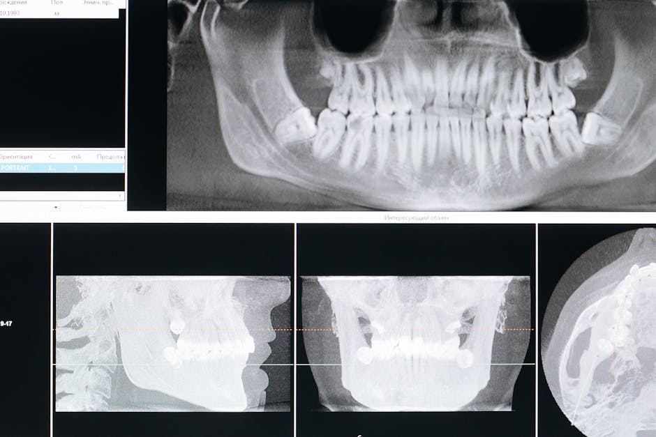
Photo by cottonbro studio on Pexels
These refer to having too many or too few teeth.
- Anodontia/Hypodontia/Oligodontia: These are degrees of missing teeth.
- Anodontia: Complete absence of all teeth (very rare).
- Hypodontia: Absence of one or a few teeth (common, often wisdom teeth, lateral incisors, or premolars).
- Oligodontia: Absence of six or more teeth.
- Cause: Often genetic, sometimes associated with syndromes like ectodermal dysplasia.
- Supernumerary Teeth (Hyperdontia): Having extra teeth.
- Mesiodens: Most common supernumerary tooth, located between the maxillary central incisors.
- Paramolar: Supernumerary tooth buccal or lingual to a molar.
- Distomolar/Fourth Molar: Supernumerary tooth distal to a third molar.
- Clinical Impact: Can cause crowding, impaction of normal teeth, or eruption problems.
Tooth Size Anomalies

Photo by Towfiqu barbhuiya on Pexels
These relate to teeth being unusually large or small.
- Microdontia: Teeth are smaller than normal.
- Generalized Microdontia: All teeth are small (rare, e.g., pituitary dwarfism).
- Relative Generalized Microdontia: Normal-sized teeth appear small in a large jaw.
- Localized Microdontia: Specific teeth are small (common, e.g., "peg lateral" incisor, third molars).
- Macrodontia: Teeth are larger than normal.
- Generalized Macrodontia: All teeth are large (rare, e.g., pituitary gigantism).
- Relative Generalized Macrodontia: Large teeth appear normal in a large jaw.
- Localized Macrodontia: Specific teeth are large (uncommon).
Tooth Shape Anomalies
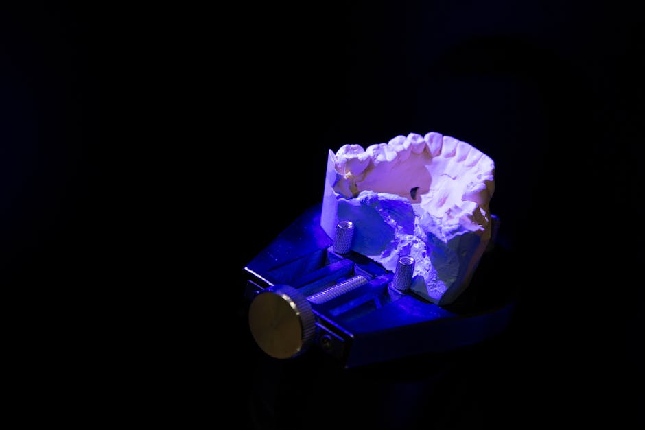
Photo by Ivan Babydov on Pexels
These are structural deviations from the typical tooth form.
- Gemination (Twinning): A single tooth bud attempts to divide, resulting in a tooth with an unusually wide crown that appears to be two teeth but has a single root canal. You'll count a normal number of teeth in the arch.
- Fusion: Union of two separate tooth buds, resulting in a large, irregularly shaped tooth with two distinct root canals. You'll count one less tooth than normal in the arch.
- Concrescence: Union of two teeth by cementum only, usually occurring after tooth formation is complete. Most common with maxillary molars.
- Dilaceration: An abnormal bend or curve in the root or crown of a tooth. Often caused by trauma during tooth development.
- Dens invaginatus (Dens in dente): "Tooth within a tooth." An invagination of the enamel organ into the dental papilla before calcification. Creates a deep pit or crevice, often on maxillary lateral incisors, predisposing to decay.
- Dens evaginatus: An accessory cusp or tubercle on the occlusal surface, usually in premolars. Contains pulp tissue and can be prone to fracture.
- Taurodontism: Teeth with elongated pulp chambers and short roots. Associated with some syndromes.
- Enamel Pearl (Enameloma): Small, spherical projection of enamel located on the root surface, usually near the furcation of molars. Can contribute to periodontal problems.
Tooth Structure Anomalies

Photo by cottonbro studio on Pexels
These involve defects in the formation of enamel or dentin.
- Amelogenesis Imperfecta (AI): A group of inherited disorders affecting enamel formation. Can present as hypoplastic (thin, pitted enamel), hypocalcified (soft, chalky enamel), or hypomaturation (mottled enamel) types. Dentin and pulp are normal.
- Dentinogenesis Imperfecta (DI): An inherited disorder affecting dentin formation. Teeth appear opalescent (bluish-brown), have short roots, obliterated pulp chambers, and bell-shaped crowns. Enamel chips off easily.
- Dentin Dysplasia: Two types.
- Type I (Radicular): Normal crowns, extremely short or absent roots ("rootless teeth"), obliterated pulp chambers.
- Type II (Coronal): Normal root length, enlarged thistle-shaped pulp chambers, pulp stones, primary teeth are amber/translucent.
- Regional Odontodysplasia (Ghost Teeth): Rare, non-hereditary developmental anomaly where a localized group of teeth in one quadrant exhibits a marked reduction in radiodensity, large pulp chambers, and short roots. Enamel and dentin are very thin and poorly mineralized, giving a "ghostly" appearance.
Jaw and Facial Anomalies
While not strictly dental, these often impact oral structures.
- Cleft Lip and Palate: Failure of fusion of facial processes during development. Can range from a small notch to a complete separation of lip and palate. Causes significant dental problems (missing teeth, supernumerary teeth, malocclusion).
graph TD
    A["Developmental Anomalies"] --> B["Tooth Number"]
    A --> C["Tooth Size"]
    A --> D["Tooth Shape"]
    A --> E["Tooth Structure"]

    B --> B1["Too Few (Hypodontia, Anodontia)"]
    B --> B2["Too Many (Supernumerary)"]

    C --> C1["Small (Microdontia)"]
    C --> C2["Large (Macrodontia)"]

    D --> D1["Gemination"]
    D --> D2["Fusion"]
    D --> D3["Concrescence"]
    D --> D4["Dilaceration"]
    D --> D5["Dens Invaginatus"]
    D --> D6["Dens Evaginatus"]
    D --> D7["Taurodontism"]
    D --> D8["Enamel Pearl"]

    E --> E1["Amelogenesis Imperfecta (Enamel)"]
    E --> E2["Dentinogenesis Imperfecta (Dentin)"]
    E --> E3["Dentin Dysplasia (Dentin/Pulp)"]
    E --> E4["Regional Odontodysplasia (Enamel/Dentin)"]
3. Worked Example
Let's say a 10-year-old patient presents to your clinic with a chief complaint of "my front tooth looks weird and I can't chew on it properly." Upon examination, you notice a maxillary left lateral incisor that appears unusually small and pointed, almost conical. You also observe that the tooth has a deep pit on its lingual surface. Radiographically, you see an infolding of enamel and dentin into the pulp chamber. The patient's other teeth appear normal.
This presentation strongly suggests Localized Microdontia (specifically a "peg lateral" incisor) combined with Dens Invaginatus (Dens in Dente). The small, pointed shape is characteristic of a peg lateral, which is a form of microdontia. The deep lingual pit and radiographic infolding are classic signs of dens invaginatus. The combination makes the tooth prone to decay and potentially structural weakness, explaining the chewing discomfort.
4. Key Takeaways
- Developmental anomalies arise from genetic or environmental factors affecting tooth or tissue formation.
- Hypodontia is missing teeth, supernumerary teeth means extra teeth; both can cause alignment issues.
- Microdontia (small) and macrodontia (large) describe tooth size variations.
- Gemination is one tooth attempting to split, while fusion is two separate teeth joining.
- Dens invaginatus ("tooth within a tooth") and dens evaginatus (extra cusp) are common shape anomalies.
- Amelogenesis Imperfecta affects enamel, and Dentinogenesis Imperfecta affects dentin.
- Regional odontodysplasia causes "ghost teeth" with very poor mineralization.
- Cleft lip/palate significantly impacts oral development and dental alignment.
Common mistakes you should avoid:
- Confusing gemination (normal tooth count) with fusion (one fewer tooth count).
- Misidentifying a dens invaginatus as simply a deep pit without considering the internal structure.
- Forgetting that some anomalies are inherited (like AI, DI) and others can be multifactorial or traumatic (like dilaceration).
- Overlooking the clinical impact – these aren't just curiosities; they often need intervention.
5. Now Try It
Imagine a patient comes in with two unusually large teeth fused together in their mandibular incisor region, and when you count, it seems like they are missing one tooth in that area. Also, their primary teeth had a distinct bluish-brown translucent appearance, but their permanent teeth are only mildly affected.
What two major developmental anomalies are most likely present, and why? Describe what each anomaly means.
Success looks like: Correctly identifying Fusion and Dentinogenesis Imperfecta (Type II if considering the primary vs. permanent difference) and providing a brief, accurate definition for each based on the provided symptoms.
Frequently asked about Developmental Anomalies of Oral and Dental Structures
More from Oral pathology
Get the full Oral pathology curriculum
Clone the complete plan to your dashboard for unlimited AI-generated notes, practice quizzes, and a personalised revision schedule.
Create Free Account